Figures & data
Figure 1 Isolation of purified synapse-specific mitochondrial fraction from rat brain cortices.
Abbreviations: SDH, succinate dehydrogenase; SM, synapse-specific mitochondria; malo, malonate; PNS, post-nuclear supernatant; PMS, post-mitochondrial supernatant; CMP, crude mitochondrial pellet.
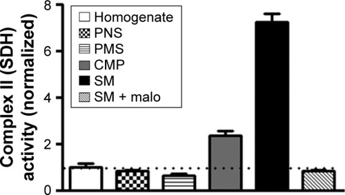
Figure 2 SM of rats developmentally exposed to Pb have increased levels of ROS and RNS and the increase is ameliorated by ascorbate.
Abbreviations: SM, synapse-specific mitochondria; Pb, lead; ROS, reactive oxygen species; RNS, reactive nitrogen species; DCFH-DA, 2′,7′-dichlorofluorescein diacetate; Ctrl, control; Asc, ascorbic acid; SEM, standard error of the mean; NOx, total nitrites and nitrates.
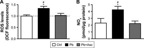
Figure 3 SM from cerebral cortices of rat pups exposed to Pb show increased oxidative damage to lipids and proteins and have reduced antioxidant capacity and their redox status is rescued by ascorbic acid supplementation.
Abbreviations: SM, synapse-specific mitochondria; Pb, lead; TBARS, thiobarbituric acid reactive substances; ABTS, 2,2′-azino-bis(3-ethylbenzothiazoline-6-sulfonic acid); Ctrl, control; Asc, ascorbic acid; SEM, standard error of the mean.
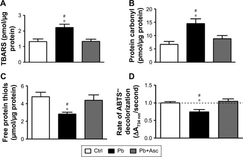
Figure 4 SM from cerebral cortices of rat pups exposed to Pb show alterations in basal mitochondrial membrane potential as well as energization-induced generation of mitochondrial membrane potential and ascorbic acid abolishes these alterations.
Abbreviations: SM, synapse-specific mitochondria; Pb, lead; MMP, mitochondrial membrane potential; Ctrl, control; Asc, ascorbic acid.
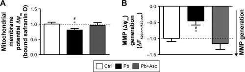
Figure 5 Pb induced alternations in the bioenergetic properties of cortical SM are mitigated by ascorbate supplementation.
Abbreviations: SM, synapse-specific mitochondria; Pb, lead; ETC, electron transport chain; Ctrl, control; Asc, ascorbic acid.
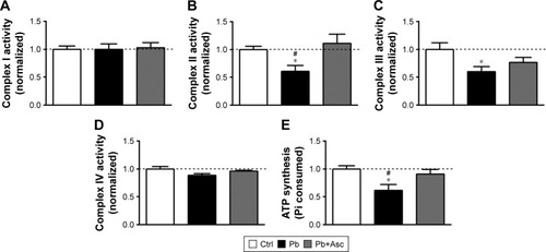
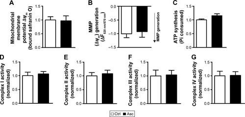
Figure S1 Supplementation of ascorbic acid alone does not affect the bioenergetics of cortical SM.
Notes: MMP at both basal states (A) and in presence of substrates (B) was not altered in SM fraction isolated from rat pups supplemented with ascorbic acid when compared to aged matched controls. Ascorbic acid supplementation did not affect other mitochondrial functions, including ATP synthesis (C) and activities of complexes I–IV (D–G). Data are represented as mean ± SEM (n=5 pups from 2 litters). Difference between the groups were assessed statistically using unpaired two-tailed Student’s t-test.
Abbreviations: SM, synapse-specific mitochondria; MMP, mitochondrial membrane potential; Ctrl, control; Asc, ascorbic acid; SEM, standard error of the mean.