
Figures & data
Figure 1 Illustration of the three types of the aortic arch. The upper line indicates the level of the top of the aortic arch, and the dotted line indicates the level of the origin of the brachiocephalic branch.
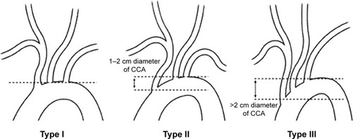
Figure 2 (A) Bovine arch – left common carotid artery originating from the innominate artery. (B) Bovine arch – common origin of the innominate artery and left common carotid artery. (C) True bovine arch.
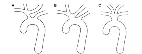
Table 1 Carotid and vertebral artery lesions of the patients compared to aortic arch types
Table 2 Carotid and vertebral artery lesions of the patients evaluated according to the presence of bovine arch
Table 3 Demographic characteristics of patients evaluated according to aortic arch types
Table 4 Carotid and vertebral artery stenosis and stenting results according to aortic arch types