
Figures & data
Table 1 The MNI coordinates of the selected ROIs in the emotion network
Table 2 Comparison of the demographic variables and psychological measurements between the Alex and the HC groups
Figure 1 Brain regions in the emotion network showing significant differences on ALFF between the Alex and the HC groups.
Notes: Red balls indicate Alex group > HC group; blue balls indicate Alex group < HC group; gray balls represent other brain regions in the emotion network.
Abbreviations: Alex, alexithymia; aIns, pOFC, anterior insula, posterior orbitofrontal cortex; ALFF, amplitude of low-frequency fluctuation; Amy vStr vGP Thal, Bi, bilateral amygdala, ventral striatum, ventral globus pallidus, thalamus; Amy_L, left basolateral amygdala; dmPFC, dorsal medial prefrontal cortex; frOP, R, right frontal operculum; HC, healthy control; Hy, hypothalamus; IFG, R, right inferior frontal gyrus; latOCC/Temp, R, right lateral occipital cortex/temporal lobe; midIns, Bi dPut, R, bilateral middle insula, right dorsal putamen; MT+, Bi, bilateral middle temporal complex; PAG/thal, periaqueductal gray/thalamus; pgACC, pregenual anterior cingulate cortex; preSMA, IFG, L, left pre-supplementary motor area, inferior frontal gyrus; rdACC, rostral dorsal anterior cingulate cortex; V4, R, right visual area 4; V8/sCB, Bi, visual area 8/bilateral superior cerebellum; vaIns/frOP/TP, RpOFC, Bi, ventral anterior insula/frontal operculum/right temporal pole, bilateral posterior orbitofrontal cortex; vaIns/TC/OFC, Bi, bilateral ventral anterior insula/temporal cortex/orbitofrontal cortex; vStr, midIns, HCMP, L, left ventral striatum, middle insula, hippocampus.
Abbreviations: Alex, alexithymia; aIns, pOFC, anterior insula, posterior orbitofrontal cortex; ALFF, amplitude of low-frequency fluctuation; Amy vStr vGP Thal, Bi, bilateral amygdala, ventral striatum, ventral globus pallidus, thalamus; Amy_L, left basolateral amygdala; dmPFC, dorsal medial prefrontal cortex; frOP, R, right frontal operculum; HC, healthy control; Hy, hypothalamus; IFG, R, right inferior frontal gyrus; latOCC/Temp, R, right lateral occipital cortex/temporal lobe; midIns, Bi dPut, R, bilateral middle insula, right dorsal putamen; MT+, Bi, bilateral middle temporal complex; PAG/thal, periaqueductal gray/thalamus; pgACC, pregenual anterior cingulate cortex; preSMA, IFG, L, left pre-supplementary motor area, inferior frontal gyrus; rdACC, rostral dorsal anterior cingulate cortex; V4, R, right visual area 4; V8/sCB, Bi, visual area 8/bilateral superior cerebellum; vaIns/frOP/TP, RpOFC, Bi, ventral anterior insula/frontal operculum/right temporal pole, bilateral posterior orbitofrontal cortex; vaIns/TC/OFC, Bi, bilateral ventral anterior insula/temporal cortex/orbitofrontal cortex; vStr, midIns, HCMP, L, left ventral striatum, middle insula, hippocampus.
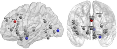
Table 3 Brain regions in the emotion network showing significant differences on ALFF between the Alex and the HC groups
Table 4 Comparison analysis of gray matter on GM density between the Alex and the HC groups
Figure 2 The functional connectivity showing significant differences between the Alex and the HC groups.
Notes: Red balls indicate Alex group > HC group; blue balls indicate Alex group < HC group and the brain regions with weak functional connectivity; gray balls represent other brain regions in the emotion network. Blue lines indicate weak functional connectivity in Alex group, compared with HC group.
Abbreviations: Alex, alexithymia; dmPFC, dorsal medial prefrontal cortex; frOP, aIns, anterior insula, frontal operculum; HC, healthy control; Hy, hypothalamus; IFG, inferior frontal gyrus; latOCC, lateral occipital cortex; MT+, middle temporal complex; PAG, periaqueductal gray; PCC, posterior cingulate cortex; PARA, paracentral lobule; pgACC, pregenual anterior cingulate cortex; pOFC, anterior insula, posterior orbitofrontal cortex; preSMA, L, left pre-supplementary motor area; STG, superior temporal gyrus; V8/sCB, visual area 8/ superior cerebellum; vaIns, ventral anterior insula; vGP, ventral globus pallidus; vStr, ventral striatum.
Abbreviations: Alex, alexithymia; dmPFC, dorsal medial prefrontal cortex; frOP, aIns, anterior insula, frontal operculum; HC, healthy control; Hy, hypothalamus; IFG, inferior frontal gyrus; latOCC, lateral occipital cortex; MT+, middle temporal complex; PAG, periaqueductal gray; PCC, posterior cingulate cortex; PARA, paracentral lobule; pgACC, pregenual anterior cingulate cortex; pOFC, anterior insula, posterior orbitofrontal cortex; preSMA, L, left pre-supplementary motor area; STG, superior temporal gyrus; V8/sCB, visual area 8/ superior cerebellum; vaIns, ventral anterior insula; vGP, ventral globus pallidus; vStr, ventral striatum.
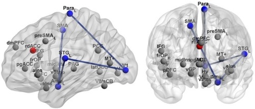
Table 5 Comparison of the functional and structural connectivity between the Alex and the HC groups