Figures & data
Figure 1 Flow diagram of the study.
Abbreviations: Erk, extracellular signal-regulated kinase; PCR, polymerase chain reaction; SAH, subarachnoid hemorrhage; TNF-α, tumor necrosis factor-alpha.
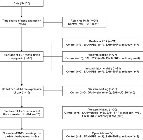
Figure 2 Changes in caspase-3, bax and bcl-2 mRNA at different time points after SAH in the hypothalamus.
Notes: (A) Caspase-3 mRNA increased 24 h after SAH and began to return to baseline at 7 days after SAH (n=6–7/group). (B) Bax mRNA and (C) bcl-2 mRNA increased 48 h after SAH (n=5–6/group). *p<0.05, **p<0.01 versus sham-operated control group.
Abbreviation: SAH, subarachnoid hemorrhage.
Abbreviation: SAH, subarachnoid hemorrhage.
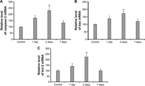
Figure 3 The effects of TNF-α antibody on the expression of caspase-3, bax and bcl-2 in the hypothalamus after SAH.
Notes: Rats were microinfused with anti-TNF-α antibody, or the same volume of PBS, 30 min before surgery. The hypothalamus was dissected out 48 h later. (A) Representative immunoblots and quantitative analyses showing the protein levels of TNF-α (n=7/group) relative to β-actin in the hypothalamus. (B) Changes in caspase-3 mRNA (n=6–7/group), bax mRNA (n=5–7/group) and bcl-2 mRNA (n=5/group). Representative immunoblots and quantitative analyses showing the protein levels of (C) caspase-3 (n=7/group), (D) bax (n=8–9/group) and (E) bcl-2 (n=6–10/group) relative to β-actin in the hypothalamus (*p<0.05, **p<0.01 versus sham-operated control group, #p<0.05 versus PBS group).
Abbreviations: SAH, subarachnoid hemorrhage; TNF-α, tumor necrosis factor-alpha.
Abbreviations: SAH, subarachnoid hemorrhage; TNF-α, tumor necrosis factor-alpha.
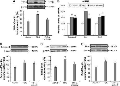
Figure 4 Immunohistochemistry showing the changes of caspase-3, bax and bcl-2 in the hypothalamus 48 h after SAH.
Notes: Rats were treated as described in . Representative coronal sections are also shown. Scale bar =100 μm.
Abbreviations: SAH, subarachnoid hemorrhage; TNF-α, tumor necrosis factor-alpha.
Abbreviations: SAH, subarachnoid hemorrhage; TNF-α, tumor necrosis factor-alpha.
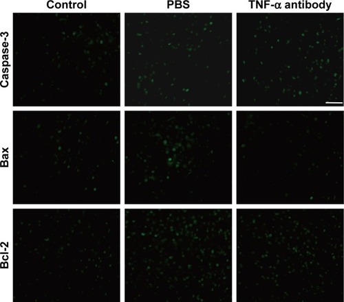
Figure 5 The effects of anti-TNF-α antibody on the levels of p-Erk in the hypothalamus 48 h after SAH.
Notes: (A) The levels of p-Erk, Erk and bax in the control group, vehicle group and U0126 group were detected by Western blotting. (B) Quantitative analyses showing the levels of p-Erk relative to total Erk, and bax relative to β-actin normalized to the sham-operated control group (n=5/group, *p<0.05 versus sham-operated control group). (C) Levels of p-Erk, Erk and bax in the control group, vehicle group, anti-TNF-α antibody group and anti-TNF-α antibody+PMA group were detected by Western blotting. (D) Quantitative analyses showing the levels of p-Erk relative to total Erk, and bax relative to β-actin normalized to sham-operated control group (n=5/group, **p<0.01 versus sham-operated control group).
Abbreviations: Erk, extracellular signal-regulated kinase; PMA, phorbol-12-myristate-13-acetate; SAH, subarachnoid hemorrhage; TNF-α, tumor necrosis factor-alpha.
Abbreviations: Erk, extracellular signal-regulated kinase; PMA, phorbol-12-myristate-13-acetate; SAH, subarachnoid hemorrhage; TNF-α, tumor necrosis factor-alpha.
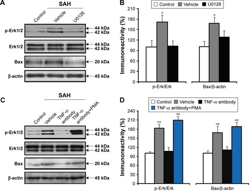
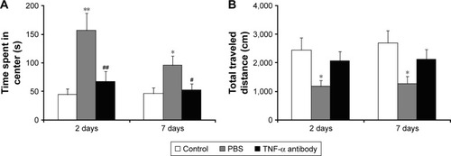
Figure 6 The effects of anti-TNF-α antibody on anxiety-like behavior.
Notes: Rats were treated as described in . Open field analysis was performed on both days 2 and 7 after SAH. (A) Time spent in the center and (B) the total distance traveled were respectively recorded (n=8/group, *p<0.05, **p<0.01 versus sham-operated control group; #p<0.05, ##p<0.01 versus PBS group).
Abbreviations: SAH, subarachnoid hemorrhage; TNF-α, tumor necrosis factor-alpha.
Abbreviations: SAH, subarachnoid hemorrhage; TNF-α, tumor necrosis factor-alpha.