
Figures & data
Figure 1 Case 1 magnetic resonance imaging and MRA of brain.
Note: (A) DWI, (B) T1FLAIR, (C) T2FLAIR, and (D) MRA. It showed infarction on left posterior horn of the lateral ventricle and the splenium of the corpus callosum (A–C, arrow) and occlusion of the posterior cerebral artery (D arrow).
Abbreviations: DWI, diffusion-weighted imaging; T1FLAIR, T1 fluid-attenuated inversion recovery imaging; T2FLAIR, T2 fluid-attenuated inversion recovery imaging; MRA, magnetic resonance angiography.
Abbreviations: DWI, diffusion-weighted imaging; T1FLAIR, T1 fluid-attenuated inversion recovery imaging; T2FLAIR, T2 fluid-attenuated inversion recovery imaging; MRA, magnetic resonance angiography.
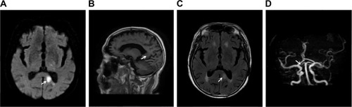
Table 1 Case 1 MoCA
Figure 2 Case 2 magnetic resonance imaging and MRA of brain.
Note: (A) DWI, (B) T2FLAIR, (C) T1FLAIR, (D) DWI, (E) T2FLAIR, (F) T1FLAIR. It showed acute infarcts in the left centrum semiovale, involving the body of corpus callosum (A–C, arrow) and multiple lacunae of infarction (D–F, arrow).
Abbreviations: DWI, diffusion-weighted imaging; T1FLAIR, T1 fluid-attenuated inversion recovery imaging; T2FLAIR, T2 fluid-attenuated inversion recovery imaging; MRA, magnetic resonance angiography.
Abbreviations: DWI, diffusion-weighted imaging; T1FLAIR, T1 fluid-attenuated inversion recovery imaging; T2FLAIR, T2 fluid-attenuated inversion recovery imaging; MRA, magnetic resonance angiography.
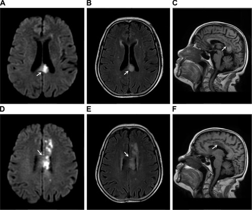
Table 2 Case 2 MoCA