
Figures & data
Figure 1 Pedigree of the family. Filled symbols, affected subjects; open symbols, unaffected; a diagonal line through a symbol, deceased; short arrow, proband; half filled symbols, mutation carrier; circles, female; squares, male. DNA available in IV2, IV3, IV4, V5, V6, V7, and V8. The proband was IV2 (arrow), a 51-year-old female with the MAPT P301L mutation presenting as bvFTD clinically. One sibling (IV4) and two children of the patient (V6 and V8) were healthy mutation carriers with the same mutation.
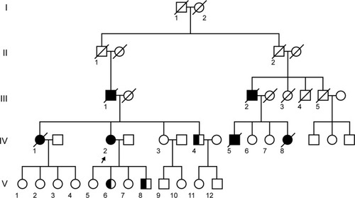
Table 1 The patient’s performance on MES scale
Figure 2 The neuroimaging of the proband and her sibling. For the proband, the T1-weighted axial MRI scan showed bilateral asymmetry of frontotemporal atrophy, more significant on the left (A). The predominantly frontotemporal hypoperfusion and hypometabolism were observed on ASL-MRI (B) and FDG-PET (C), respectively, in the proband. FDG-PET studies in her sibling (IV4, mutation carrier, indexed in ) revealed no significant abnormalities on visual inspection (D).
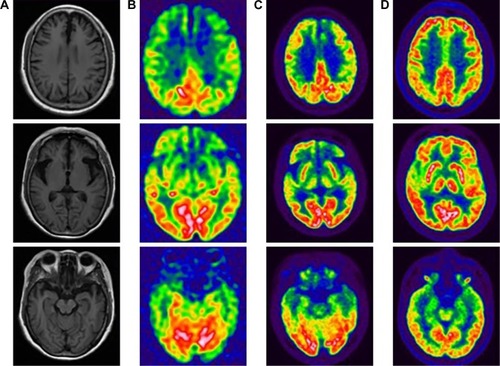
Table 2 The Chinese patients reported in the literature with MAPT mutations
Table S1 MAPT primers