Figures & data
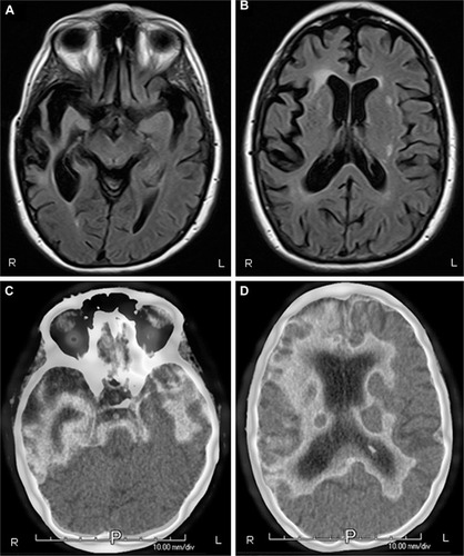
Figure 1 (A and B) MRI of brain, axial T2 Flair view, showing prominent bitemporal, right greater than left, atrophy. (C and D) PET imaging illustrating bitemporal and right frontal hypometabolism. There is extension of the hypometabolism to the right parietal lobe. All images are radiological convention with the left hemisphere on the right and the right hemisphere on the left.
Abbreviations: R, right; L, left; P, posterior; MRI, magnetic resonance imaging; PET, positron emission tomography.