Figures & data
Figure 1 Clinical course and results of immunohistochemistry, imaging, and pathology analyses.
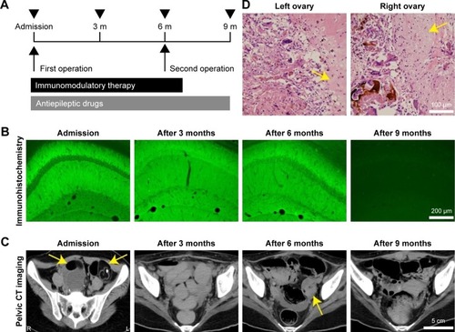
Notes: (A) Timeline of examination and therapy. Black arrow heads indicate pelvic CT scans and immunohistochemical analyses at 3-month intervals for 9 months. (B) Temporal profile of immunohistochemical analysis of the patient’s CSF. Robust reactivity in the hippocampus was observed on admission. This reactivity remained after the first operation and the immunotherapy (after 3 and 6 months). Note that no reactivity was confirmed after the second operation (after 9 months). Scale bar: 200 μm. (C) Pelvic CT scan images. Bilateral ovarian teratomas on admission (yellow arrows) were enucleated in the first operation. Six months later, recurrence of the left ovarian teratoma could be seen (yellow arrow). The bilateral ovaries were completely resected by salpingo-oophorectomy (second operation; after 9 months). Scale bar: 5 cm. (D) Pathological findings. In addition to the left ovary, the pathological examination of the right ovary showed a mature cystic teratoma, indicating bilateral recurrence of the ovarian teratomas. Yellow arrows show neuroglial tissues. Scale bar: 100 μm.
Abbreviations: CSF, cerebrospinal fluid; CT, computed tomography; m, months.
Abbreviations: CSF, cerebrospinal fluid; CT, computed tomography; m, months.