Figures & data
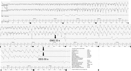
Figure 1 An inadequate response in the ninth session (bilateral brief pulse ECT, Low 0.5, 100%), in which no motor seizure was observed, and the peak heart rate did not increase from the baseline.
Notes: EEG in channels 1 and 2, EMG in channel 3, and ECG in channel 4. The standard fronto-mastoid EEG electrode placements were used. The gain of the EEG amplifiers was set at 200 μV/cm, and the gain of the EMG and ECG amplifiers was set at 1,000 μV/cm.
Abbreviations: ECG, electrocardiogram; ECT, electroconvulsive therapy; EEG, electroencephalogram; EMG, electromyogram.
Abbreviations: ECG, electrocardiogram; ECT, electroconvulsive therapy; EEG, electroencephalogram; EMG, electromyogram.
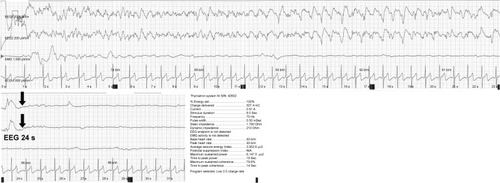
Figure 2 Successful seizure induction following the third stimulation of the eleventh session (right unilateral ultrabrief pulse ECT, Low 0.25, 15%), in which a tonic-clonic motor seizure was first observed.
Notes: EEG in channels 1 and 2, EMG in channel 3, and ECG in channel 4. The standard frontomastoid EEG electrode placements were used. The gain of the EEG amplifiers was set at 200 μV/cm, and the gain of the EMG and ECG amplifiers was set at 1,000 μV/cm.
Abbreviations: ECG, electrocardiogram; ECT, electroconvulsive therapy; EEG, electroencephalogram; EMG, electromyogram.
Abbreviations: ECG, electrocardiogram; ECT, electroconvulsive therapy; EEG, electroencephalogram; EMG, electromyogram.
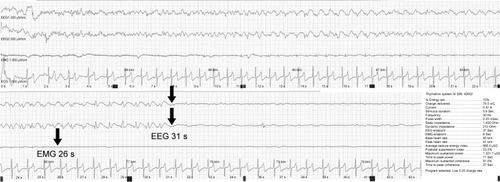
Figure 3 An adequate therapeutic seizure in the 16th session (right unilateral ultrabrief pulse ECT, Low 0.25, 100%), which was associated with high-amplitude slow waves and postictal suppression on the ictal electroencephalogram.
Notes: EEG in channels 1 and 2, EMG in channel 3, and ECG in channel 4. The standard frontomastoid EEG electrode placements were used. The gain of the EEG amplifiers was set at 200 μV/cm, and the gain of the EMG and ECG amplifiers was set at 1,000 μV/cm.
Abbreviations: ECG, electrocardiogram; ECT, electroconvulsive therapy; EEG, electroencephalogram; EMG, electromyogram.
Abbreviations: ECG, electrocardiogram; ECT, electroconvulsive therapy; EEG, electroencephalogram; EMG, electromyogram.