Figures & data
Figure 1 Diminished expression of TH in substantia nigra in PD and LID rats.
Notes: Western blot for TH of extracts from substantia nigra of sham (1), PD (2) and LID (3) rats. The optical density was quantified by densitometry, and the value of TH is expressed as the percentage of GAPDH (TH/GAPDH × 100% ± SD). #p < 0.01 versus sham.
Abbreviations: LID, levodopa-induced dyskinesia; PD, Parkinson’s disease; TH, tyrosine hydroxylase.
Abbreviations: LID, levodopa-induced dyskinesia; PD, Parkinson’s disease; TH, tyrosine hydroxylase.
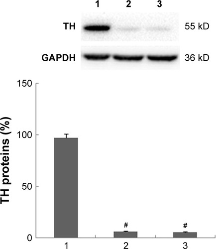
Figure 2 PD rats show different AIMs scores after the administration of l-DOPA, D1R antagonist or agonist.
Notes: Axial (A), limb (B), orolingual (C) and locomotor (D). AIMs scores were observed at 2, 7, 12, 17 and 22 days. Increased AIMs scores in PD rats were observed after the administration of l-DOPA. Meanwhile, the group treated with l-DOPA and D1R agonist showed increasing AIMs scores; however, rats treated with D1R antagonist, SCH23390, plus l-DOPA showed no increasing AIMs scores compared to rats peritoneally injected with l-DOPA alone. Data are presented as mean ± SD. #p < 0.05, versus day 2; *p < 0.05, versus l-DOPA group. Data are statistically analyzed by one-way ANOVA test.
Abbreviations: AIMs, abnormal involuntary movements; ANOVA, analysis of variance; d, days; D1R, D1 dopamine receptor; l-DOPA, L-3,4-dihydroxyphenylalanine; PD, Parkinson’s disease.
Abbreviations: AIMs, abnormal involuntary movements; ANOVA, analysis of variance; d, days; D1R, D1 dopamine receptor; l-DOPA, L-3,4-dihydroxyphenylalanine; PD, Parkinson’s disease.
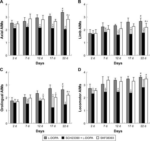
Figure 3 D1R interacts with Shp-2 in the striatal neurons.
Notes: Striatal proteins were coimmunoprecipitated with anti-D1R and anti-Shp-2 antibodies (A and B). Representative immunoblots showing D1R and Shp-2 interactions in striatal neurons as detected by coimmunoprecipitation. No precipitating antibody, an irrelevant IgG was used in L2, L3, respectively. The D1R antibody (L4A) or Shp-2 antibody (L4B) was used in L4 for normal rats and L5 for LID rats, respectively. No striatal proteins and antibodies were used in L1.
Abbreviations: IP, immunoprecipitation; D1R, D1 dopamine receptor; L1, lane 1; L2, lane 2; L3, lane 3; L4, lane 4; L5, lane 5; LID, levodopa-induced dyskinesia.
Abbreviations: IP, immunoprecipitation; D1R, D1 dopamine receptor; L1, lane 1; L2, lane 2; L3, lane 3; L4, lane 4; L5, lane 5; LID, levodopa-induced dyskinesia.
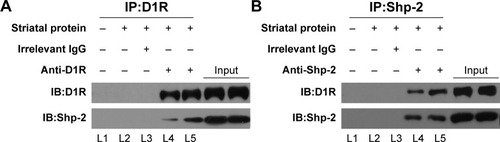
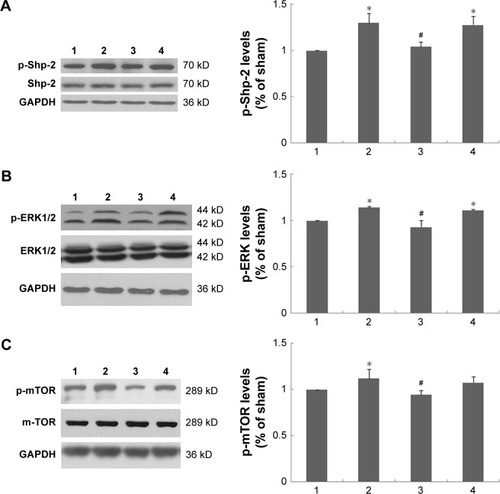
Figure 4 Molecular events underlying LID involving D1R/Shp-2 complex and its downstream signaling factors, such as ERK1/2 and mTOR.
Notes: The bands represent immunoblot images detected by antibodies against p-Shp-2 (A), p-ERK (B) and p-mTOR (C). Proteins were analyzed from the sham group (1), l-DOPA group (2), SCH23390 + l-DOPA group (3), SKF38393 group (4). Repeated administration of l-DOPA increased the level of p-Shp-2, p-ERK1/2 and p-mTOR. SKF38393 increased the levels similarly. Conversely, SCH23390 plus L-DOPA prevented the increase. Data are presented as mean ± SD. *p < 0.05, versus sham group; #p < 0.05, versus l-DOPA group. Data are statistically analyzed by one-way ANOVA test.
Abbreviations: ANOVA, analysis of variance; D1R, D1 dopamine receptor; ERK1/2, extracellular signal-regulated kinases 1 and 2; l-DOPA, l-3,4-dihydroxyphenylalanine; LID, levodopa-induced dyskinesia.
Abbreviations: ANOVA, analysis of variance; D1R, D1 dopamine receptor; ERK1/2, extracellular signal-regulated kinases 1 and 2; l-DOPA, l-3,4-dihydroxyphenylalanine; LID, levodopa-induced dyskinesia.