Figures & data
Figure 1 The experimental flowchart.
Abbreviations: Dex, dexmedetomidine; DMSO, dimethyl sulfoxide; i.c., intracerebroventricularly; i.p., intraperitoneally; TEM, transmission electron microscopy; TUNEL, terminal deoxynucleotidyl transferase-mediated dUTP nick end labeling.
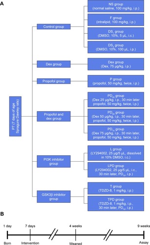
Figure 2 Injection of young rats with Dex and/or propofol did not affect their body weights in adult age.
Abbreviations: Dex, dexmedetomidine; DMSO, dimethyl sulfoxide; SEM, standard error of the mean.
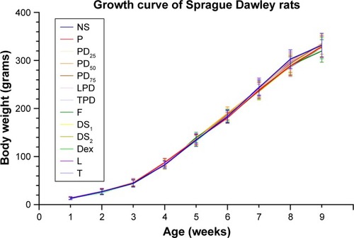
Figure 3 Pretreatment of rats with Dex at young age improved the propofol-induced impairment in spatial learning and memory ability in adult age.
Abbreviations: Dex, dexmedetomidine; DMSO, dimethyl sulfoxide; MWM, Morris water maze; SEM, standard error of the mean.
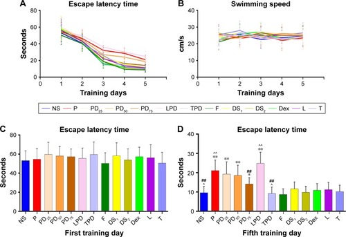
Table 1 Injection with Dex and/or propofol did not affect arterial blood gas
Figure 4 Pretreatment of young rats with Dex decreased the propofol-induced neuroapoptosis in the hippocampus in adult age.
Abbreviations: DAPI, 4′,6-diamidino-2-phenylindole; Dex, dexmedetomidine; DMSO, dimethyl sulfoxide; FITC, fluorescein isothiocyanate; SEM, standard error of the mean; TUNEL, terminal deoxynucleotidyl transferase-mediated dUTP nick end labeling.
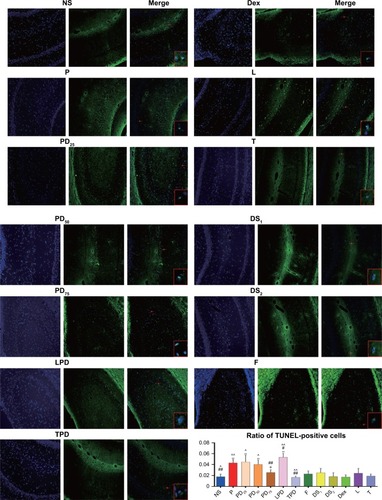
Figure 5 Pretreatment of young rats with Dex attenuated the propofol-induced long-term neurotoxicity and increased the levels of PSD95 expression in the hippocampus in adult age.
Abbreviations: DAB, 3,3′-diaminobenzidine tetrahydrochloride; Dex, dexmedetomidine; DMSO, dimethyl sulfoxide; HRP, horseradish peroxidase; SEM, standard error of the mean.
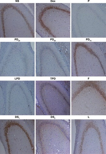
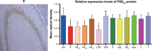
Figure 6 Western blot analysis of the relative levels of PSD95 expression in the hippocampus of rats.
Abbreviations: DMSO, dimethyl sulfoxide; sSEM, standard error of the mean.
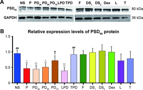
Figure 7 The ultrastructure of hippocampal neurons as revealed by TEM.
Abbreviations: DMSO, dimethyl sulfoxide; TEM, transmission electron microscopy.
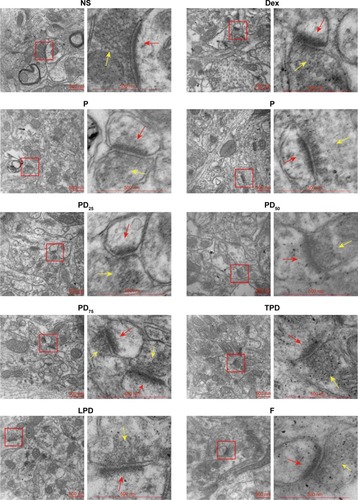
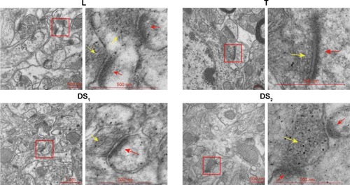
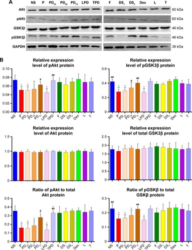
Figure 8 Pretreatment of young rats with Dex mitigated the propofol-attenuated Akt activation in the hippocampus of rats.
Abbreviations: Dex, dexmedetomidine; DMSO, dimethyl sulfoxide; SEM, standard error of the mean.