Figures & data
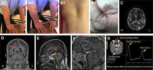
Figure 1 Signs observed in the current case.
Notes: (A1, A2) Congenital hyper-flexibility type EDS (red arrows, elbows);Citation4,Citation5 (B1, B2) recurrent urticaria and eczema on torso (B1) and hands (B2);Citation4,Citation5 (C) MS and MDD-related ventricular enlargement and occipital lobe bending (OLB, blue and white arrows, respectively);Citation1,Citation2,Citation6 OLB is also shown in (G). (D) EDS-related unilateral periventricular heterotopia (PH, red arrow), which supports the patient’s dyslexia diagnosis.Citation7 (E) Red arrow, MS lesions along body and splenum of corpus callosum. They indicate difficulties in coordinating R/L hands and may portend cognitive decline.Citation8,Citation9 Yellow arrow – craniopharyngioma (0.9 cm3) comprising papillary (soft, anterior), and adamantinous (hard, posterior) cysts bound by a single epithelium.Citation10–Citation12 (F) Yellow arrow, conjoined craniopharyngioma cysts (soft ~2.3 cm3 and hard ~0.5 cm3). At this stage, the tumor pressed against his optic chiasm (thin gray arch above the cyst, blue arrow) and caused bitemporal hemianopsia (bilateral peripheral vision losses).Citation10–Citation12 (G) Red arrow, EDS-related nasal cartilage defect.Citation4,Citation5 White arrow, OLB.Citation6 Yellow arrows, single voxel (box) MRS of papillary cyst showing characteristic alkyl fats and hydroxyl resonances at δ ≅1–2 and 3.5–4.2 ppm, respectively. Other CNS metabolites are not seen in papillary craniopharyngioma tumors. These MR-spectral characteristics are only seen in papillary craniopharyngiomas.Citation11,Citation12 Adamantinous cysts do not display MR-spectral resonances. Note, in this case report the corresponding author (NDS) is the patient.
Abbreviations: EDS, Ehlers Danlos Syndrome; MS, multiple sclerosis; MDD, major depressive disorder; OLB, occipital lobe bending; R, right; L, left.
Abbreviations: EDS, Ehlers Danlos Syndrome; MS, multiple sclerosis; MDD, major depressive disorder; OLB, occipital lobe bending; R, right; L, left.