Figures & data
Table 1 Medication administration and CIWA
Figure 1 Continuous 17-hour video EEG monitoring.
Notes: The EEG was collected digitally and stored in a computer that has seizure detection as well as spike detection software. The patient can activate a push-button event as well as the nurse to indicate if a clinical event has occurred. With the patient awake, the background showed no clear dominant occipital rhythm over the left hemisphere. There was low voltage beta activity recorded diffusely. There is irregular polymorphic Δ/θ activity recorded at 3–5 Hz maximal over the left temporoparietal region.
Abbreviation: EEG, electroencephalogram.
Abbreviation: EEG, electroencephalogram.
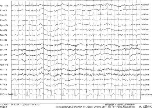
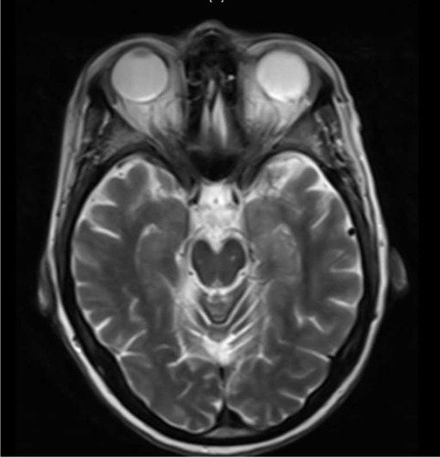
Figure 2 Current admission shows less pronounced left mesial temporal abnormality.
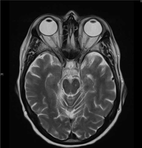
Figure 3 Admission 9 months prior showing nonspecific FLAIR abnormality in the left mesial temporal lobe.