Figures & data
Figure 1 Animal behavior tests – SPT.
Notes: (A) SP (%) in the OFT. (B) Immobility time (seconds) in the FST. (C) Total distance covered in the OFT (cm). (E) Number of entries into center zone. (D) Number of rears in the vertical direction in the EPM test. (F) Total distance covered in the EPM test (cm). (G) Time spent in open arms (seconds). (H) Number of entries into open arms in the EPM test. All data are presented as mean ± SEM (n=11–12 rats/group). Statistical analysis was conducted using ANOVA using post hoc comparisons with Tukey’s test. *P<0.05, **P<0.01, and ***P<0.001.
Abbreviations: EPM, elevated plus maze; FST, forced swim test; OFT, open field test; SEM, standard error of the mean; SP, sucrose preference; SPT, sucrose preference test.
Abbreviations: EPM, elevated plus maze; FST, forced swim test; OFT, open field test; SEM, standard error of the mean; SP, sucrose preference; SPT, sucrose preference test.
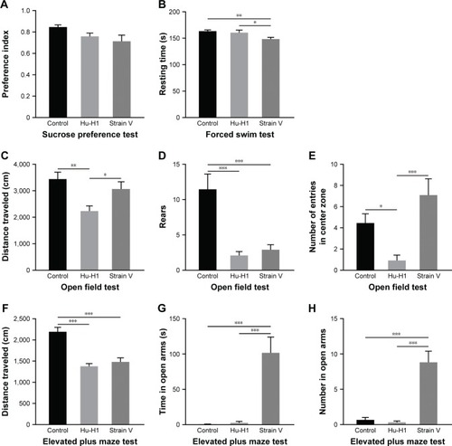
Figure 2 The average moving speed of three groups of rats in OFT.
Notes: No significance was found among the three groups. All data are presented as mean ± SEM. Statistical analysis was performed using ANOVA for post hoc comparisons with Tukey’s test.
Abbreviations: OFT, open field test; SEM, standard error of the mean.
Abbreviations: OFT, open field test; SEM, standard error of the mean.
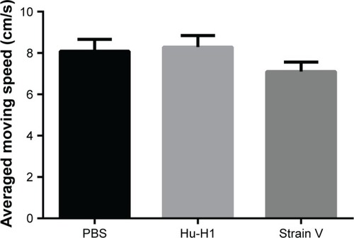
Figure 3 Immunofluorescence analysis of primary hippocampal neurons infected with BDV on day 9 postinfection.
Notes: (A) Purity of neurons stained with MAP2 followed by Cy3-labeled goat anti-rabbit IgG antibody (red). (B) BDV Hu-H1- or Strain V-infected cells combined with P24 antibody followed by FITC-labeled goat anti-mouse IgG antibody (green). (C) Nuclei of cells or DNA released from apoptotic cells were stained by DAPI (blue). (D) Merged images (magnification: 100×).
Abbreviation: BDV, Borna disease virus.
Abbreviation: BDV, Borna disease virus.
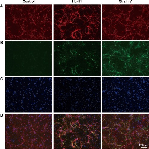
Figure 4 Identification of BDV infection in rats. BDV-specific antibodies (P24 and P40) identified the stable expression of virus in the hippocampus of rats 8 weeks postinfection compared with the control group.
Abbreviation: BDV, Borna disease virus.
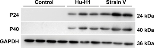
Figure 5 Western blotting of rat hippocampi.
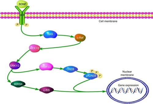
Notes: Eight selected antibodies were included: p-c-Raf, p-90RSK, p-MSK, p-ERK1/2, p-MEK1/2, p-CREB, CREB, and BDNF. Expression levels of (A) BDNF (n=7), (C) p-MEK1/2 (n=6), (G) CREB (n=4), and (H) p-CREB (n=7) were not significantly affected by BDV infection. (B) Expression level of p-c-Raf was upregulated in Strain V-infected rats compared with control rats (P<0.05, n=4). (D) Expression level of p-ERK1/2 was significantly increased in BDV-infected rats relative to control rats (P<0.001, n=7). (E) Expression level of p-MSK was significantly upregulated in Hu-H1-infected rats compared with control rats (P<0.05, n=6). (F) Expression level of p-90RSK was elevated in Hu-H1-infected rats compared with control rats (P<0.05, n=5). Bars indicate mean ± SEM. Statistical analysis was performed using ANOVA. *P<0.05 and ***P<0.001.
Abbreviations: BDV, Borna disease virus; SEM, standard error of the mean.
Abbreviations: BDV, Borna disease virus; SEM, standard error of the mean.
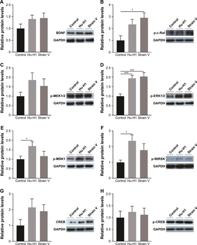
Figure 6 Western blotting of primary hippocampal neuron lysates cultured for 9 days.
Notes: (A): Identification of BDV infection in cells (A) No significant differences were found in the expression of (B) BDNF, (D) p-MEK1/2, (E) p-ERK1/2, (G) p-90RSK, and (H) p-CREB. (C) Expression level of p-c-Raf was upregulated in Hu-H1-infected neurons compared with control neurons (P<0.05). (F) Expression of p-MSK was significantly downregulated in Hu-H1-infected and Strain V-infected cells compared with mock-infected neurons (P<0.05). The experiments were repeated thrice. Values were expressed as mean ± SEM. *P<0.05.
Abbreviations: BDV, Borna disease virus; SEM, standard error of the mean.
Abbreviations: BDV, Borna disease virus; SEM, standard error of the mean.
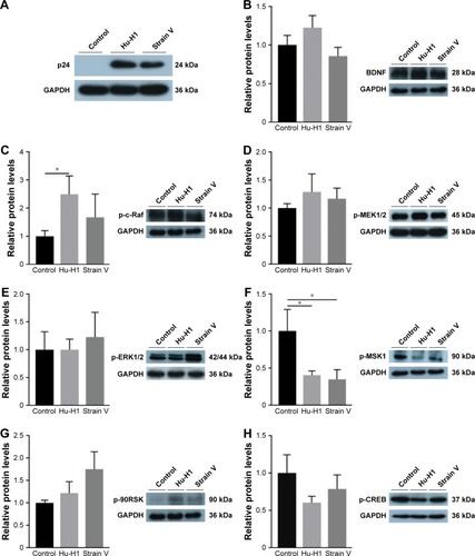
Figure 7 The ERK/CREB/BDNF cascade in this study.