Figures & data
Figure 1 Hemin induced dose-dependent necrosis and neurotoxicity in HT22 cells.
Notes: (A) Representative PI and Hoechst staining images of HT22 cells treated with hemin for 24 hours. (B) Necrotic cell death in HT22 was quantified by percentage of PI-positive cells (PI+/Hoechst+ cells). (C) The hemin neurotoxicity was confirmed by cell viability determined using CellTiter-Glo assay. The data were normalized to control group (100%). Data are expressed as mean ± SEM. Data were obtained from three independent experiments.
Abbreviation: PI, propidium iodide.
Abbreviation: PI, propidium iodide.
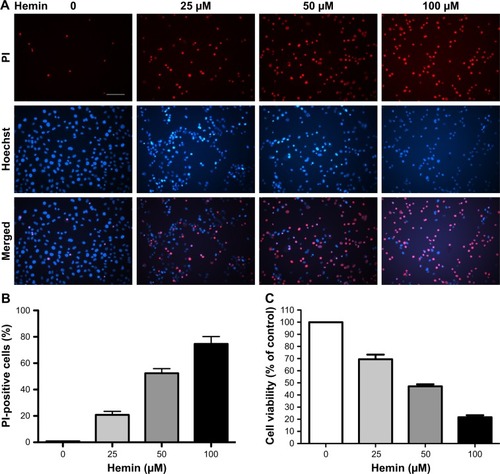
Figure 2 Cell death induced by hemin in HT22 cells was necroptosis. HT22 cells were treated with 50 μM hemin with or without 20 μM z-VAD-fmk in the presence or absence of 30 μM Nec-1 for 24 hours.
Notes: (A) Representative PI and Hoechst staining images (×200) from each treatment group. Scale bar =50 μm. (B) Quantification of the PI+ cells in each group. Results showed that Nec-1 rather than z-VAD-fmk greatly inhibited PI+ cells induced by hemin in HT22 cells. ***P<0.001 vs DMSO group; ##P<0.01 vs hemin group; &&&P<0.001 vs hemin + z-VAD-fmk group. (C) Cell viability determined using CellTiter-Glo assay further confirmed the protection by Nec-1 against hemin-induced neurotoxicity. The data were normalized to DMSO group (100%). ***P<0.001 vs DMSO group; #P<0.05 vs hemin group; &P<0.05 vs hemin + z-VAD-fmk group. Data were obtained from three independent experiments.
Abbreviations: DMSO, dimethyl sulfoxide; Nec-1, necrostatin-1; PI, propidium iodide; z-VAD-fmk, Benzyloxycarbonyl-Val-Ala-Asp(OMe)-fluoromethylketone.
Abbreviations: DMSO, dimethyl sulfoxide; Nec-1, necrostatin-1; PI, propidium iodide; z-VAD-fmk, Benzyloxycarbonyl-Val-Ala-Asp(OMe)-fluoromethylketone.
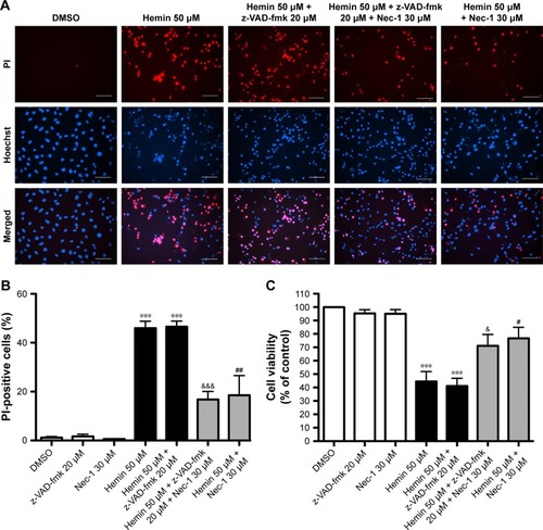
Figure 3 ROS accumulation induced by hemin mediated necrotic cell death in HT22 cells.
Notes: (A) Representative images (×400) of MitoSox Red and Hoechst fluorescence in HT22 cells at 24 hours after treatment with DMSO, hemin, and hemin + Nec-1. Results showed that ROS were robustly increased in hemin-treated HT22 cells but were significantly inhibited by Nec-1 treatment. Scale bar =50 μm. (B) HT22 cells were treated with 50 μM hemin in the presence or absence of 100 μM BHA for 24 hours. Quantification of the PI+ cells showed the protective effects of BHA on hemin-induced necrotic cell death. ***P<0.001 vs DMSO group; ###P<0.001 vs hemin group. Data were obtained from three independent experiments.
Abbreviations: BHA, butylated hydroxyanisole; DMSO, dimethyl sulfoxide; Nec-1, necrostatin-1; PI, propidium iodide.
Abbreviations: BHA, butylated hydroxyanisole; DMSO, dimethyl sulfoxide; Nec-1, necrostatin-1; PI, propidium iodide.
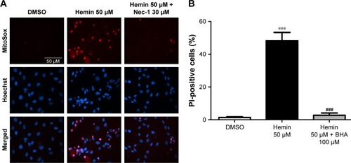
Figure 4 The levels of RIP3 protein expression were determined by Western blotting after siRNA transfection in HT22 cells.
Notes: (A) Representative Western blots showed the effect of RIP3 knockdown. (B) Densitometric quantification of RIP3 showed that RIP3 protein was greatly inhibited by RIP3 siRNA. β-Actin was used as internal control. ##P<0.01 vs negative siRNA group.
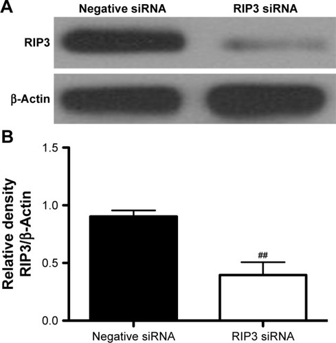
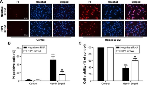
Figure 5 siRNA silencing of RIP3 attenuated hemin-induced necrotic cell death in HT22 cells.
Notes: (A) Representative PI and Hoechst staining photographs (×200) in each treatment group. Scale bar =50 μm. (B) PI+ cells were quantitated by fluorescence microscopy. Results showed that RIP3 knockdown significantly inhibited PI+ cells induced by hemin in HT22 cells. ***P<0.001 vs control group; ##P<0.01 vs hemin + negative siRNA group. (C) Cell viability was determined by CellTiter-Glo assay. Results further confirmed that RIP3 siRNA significantly inhibited neurotoxicity in HT22 cells after hemin treatment. The data were normalized to control group (100%). ***P<0.001 vs control group; ##P<0.01 vs hemin + negative siRNA group. Data were obtained from three independent experiments.
Abbreviation: PI, propidium iodide.
Abbreviation: PI, propidium iodide.