Figures & data
Figure 1 Schematic of extracorporeal blood Aβ removal system.
Note: Rapid removal of blood Aβ reduces Aβ concentrations in the blood, which might accelerate Aβ transport from the brain into the blood.
Abbreviations: Aβ, amyloid-β protein; AD, Alzheimer’s disease; conc., concentration; MCI, mild cognitive impairment.
Abbreviations: Aβ, amyloid-β protein; AD, Alzheimer’s disease; conc., concentration; MCI, mild cognitive impairment.
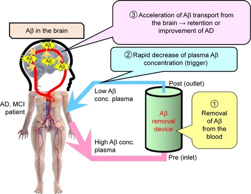
Figure 2 Images from the magnetic resonance imaging at the hemodialysis initiation.
Notes: Left, coronal; middle, axial; right, sagittal.
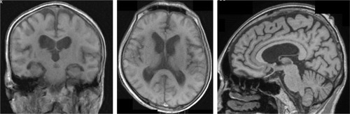
Table 1 PiB/PET evaluations of visits 1–3
Figure 3 The SUVRWCb image from the surface projection of PiB/PET.
Notes: Top, visit 1 (at the hemodialysis initiation); middle, visit 2 (6 months after the hemodialysis initiation); bottom, visit 3 (12 months after the hemodialysis initiation).
Abbreviations: PiB/PET, positron emission tomography with Pittsburgh compound B as probe; SUVRWCb, standard uptake value ratio with whole cerebellum as reference.
Abbreviations: PiB/PET, positron emission tomography with Pittsburgh compound B as probe; SUVRWCb, standard uptake value ratio with whole cerebellum as reference.
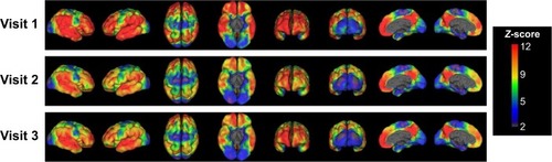
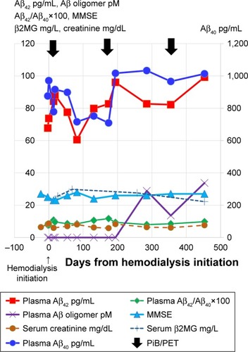
Figure 4 The concentration change of Aβs in the blood and MMSE score after the hemodialysis initiation.
Notes: The left vertical axis represents Aβ1–42 (pg/mL), Aβ oligomers (pmol/L), Aβ1–42:Aβ1–40 ratio (×100), MMSE scores, β2MG (mg/L), and creatinine (mg/dL). The right vertical axis represents Aβ1–40 (pg/mL). Black arrows indicate the day of PiB/PET. The horizontal axis represents days from the hemodialysis initiation. The blood samples were collected just before the start of each hemodialysis session for the period after the hemodialysis initiation.
Abbreviations: Aβ, amyloid-β; β2MG, β2-microglobulin; MMSE, Mini-Mental State Examination; PiB/PET, positron emission tomography with Pittsburgh compound B as probe.
Abbreviations: Aβ, amyloid-β; β2MG, β2-microglobulin; MMSE, Mini-Mental State Examination; PiB/PET, positron emission tomography with Pittsburgh compound B as probe.