Figures & data
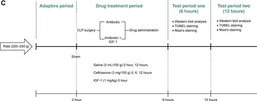
Figure 1 IGF-1 cannot improve the survival rate of septic rats.
Abbreviation: IGF-1, insulin-like growth factor-1.
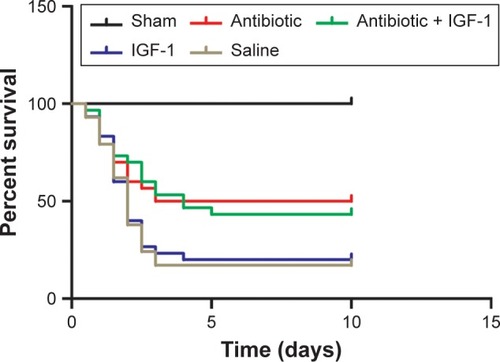
Figure 2 IGF-1 can improve the memory in a new environment of septic rats.
Abbreviation: IGF-1, insulin-like growth factor-1.
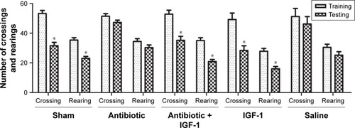
Figure 3 IGF-1 can improve the spatial learning and memory of septic rats.
Abbreviation: IGF-1, insulin-like growth factor-1.

Figure 4 IGF-1 can relieve the impairment of noxious memory in septic rats.
Abbreviation: IGF-1, insulin-like growth factor-1.
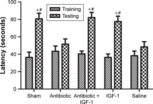
Figure 5 IGF-1 could not improve the depressive-like symptoms which might be caused by SE.
Abbreviations: CLP, cecal ligation and puncture; IG F-1, insulin-like growth factor-1; SE, septic encephalopathy.
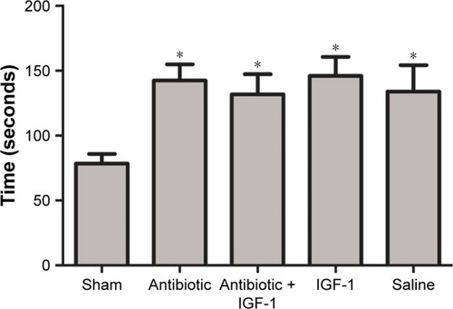
Figure 6 IGF-1 cannot improve the memory of septic rats in a new environment when it is administrated at 12, 24, or 36 hours after CLP.
Abbreviations: CLP, cecal ligation and puncture; IGF-1, insulin-like growth factor-1.
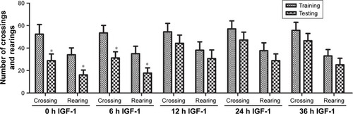
Figure 7 IGF-1 cannot improve the spatial learning and memory of septic rats when it is administrated at 12, 24, or 36 hours after CLP.
Abbreviations: CLP, cecal ligation and puncture; IGF-1, insulin-like growth factor-1.
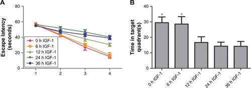
Figure 8 IGF-1 cannot improve the impairment of noxious memory of septic rats when it is administrated at 12, 24, or 36 hours after CLP.
Abbreviations: CLP, cecal ligation and puncture; IGF-1, insulin-like growth factor-1.
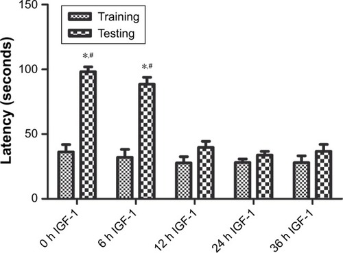
Figure 9 Inhibition of cell apoptosis in the hippocampus is associated with improvement of memory and learning during sepsis.
Abbreviation: IGF-1, insulin-like growth factor-1.
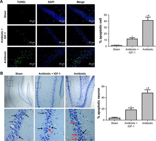
Figure 10 Cytochorme C and TNFR are activated at different time points and IGF-1 can inhibit their expression.
Abbreviations: Caspase, cysteinyl aspartate specific proteinase; CLP, cecal ligation and puncture; Cyt C, cytochrome C; IGF-1, Insulin-like growth factor-1; TNFR, tumor necrosis factor receptor.
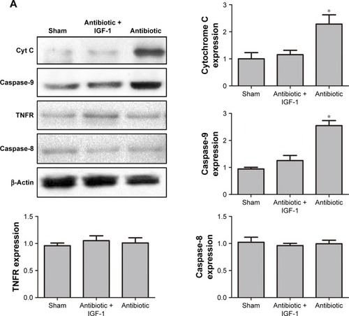
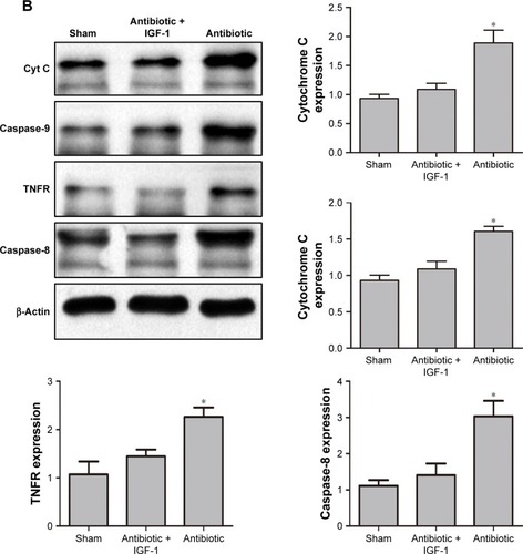
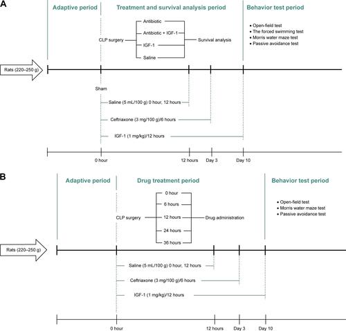
Figure S1 Experimental procedures.
Notes: (A) Experimental procedure for the first part. IGF-1, ceftriaxone, and saline were given and different duration and behavior tests were conducted 10 days after CLP. (B) Experimental procedure for the second part. IGF-1 was given at 0, 6, 12, 24, or 36 hours after surgery in each group, respectively, and then supplied every 12 hours for 10 days. Ceftriaxone and saline were given the same way as that in the first part and then behavior tests were conducted 10 days after CLP. (C) Experimental procedure for the third part. IGF-1, ceftriaxone, and saline were given at different time points. Western blot, TUNEL staining, and Nissl’s staining were conducted at 6 and 12 hours after surgery.
Abbreviations: CLP, cecal ligation and puncture; IGF-1, insulin-like growth factor-1.