
Figures & data
Figure 1 Anti-NMDA receptor antibodies in patient’s cerebrospinal fluid and serum. Anti-NMDA receptor antibodies were positive in cerebrospinal fluid (IgG, 1:32) (A) and serum (IgG, 1:320) (B) before immunotherapy. The level of anti-NMDA receptor antibodies significantly decreased in cerebrospinal fluid (IgG, 1:3.2) (C) and serum (IgG, 1:32) (D) after 5 months of immunotherapy.
Abbreviation: NMDA, N-methyl-D-aspartate.
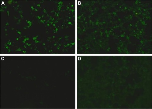
Table 1 Clinical similarities, differences, and diagnostic features in anti-NMDA receptor encephalitis and NMS