Figures & data
Table 1 Sex, Age, And Course Of Disease Between MSA And PD Groups
Table 2 EAS-EMG Results Between MSA And PD Groups
Figure 1 EAS-EMG and US-EMG of patients with MSA.
Notes: (A) Prolonged MUP duration and increased percentage of polyphasic MUPs were detected by EAS-EMG in MSA group. (B) Prolonged MUP duration was detected by US-EMG in MSA group.
Abbreviations: EAS-EMG, external anal-sphincter electromyography; US-EMG, urethral-sphincter electromyography; MSA, multiple-system atrophy; MUPs, motor unit potentials.
Abbreviations: EAS-EMG, external anal-sphincter electromyography; US-EMG, urethral-sphincter electromyography; MSA, multiple-system atrophy; MUPs, motor unit potentials.
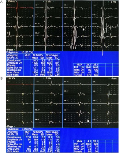
Table 3 US-EMG Results Between MSA And PD Groups
Table 4 Comparison Of Indices Obtained In EAS-EMG And US-EMG Of MSA Group
Table 5 Cutoff Points, Area Under ROC curve, Sensitivity, Specificity, And 95% CIs Of Some Parameters In EAS-EMG And US-EMG For Differentiating MSA From PD
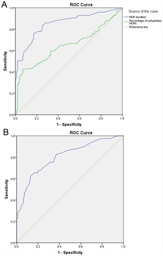
Figure 2 ROC curves for MSA and PD in EAS-EMG and US-EMG.
Notes: (A) ROC curve for differential diagnosis of MSA with PD according to mean MUP duration and percentage of polyphasic MUPs in EAS-EMG. (B) ROC curve for differential diagnosis of MSA with PD according to mean MUP duration in US-EMG.
Abbreviations: ROC, receiver-operating characteristic; EAS-EMG, external anal-sphincter electromyography; US-EMG, urethral-sphincter electromyography; MSA, multiple-system atrophy; PD, Parkinson’s disease; MUPs, motor unit potentials.
Abbreviations: ROC, receiver-operating characteristic; EAS-EMG, external anal-sphincter electromyography; US-EMG, urethral-sphincter electromyography; MSA, multiple-system atrophy; PD, Parkinson’s disease; MUPs, motor unit potentials.