Figures & data
Figure 1 (A) Chemical structure of stigmasterol (C29H48O, molecular weight: 412.69) and (B) schematic diagram of the experimental protocol. Vehicle (0.1% DMSO in 1% hydroxylethyl cellulose) and stigmasterol (20, 40, and 80 mg/kg) were given via i.p. after 2-hr ischemia. 3-MA (300 μg/kg) was given via i.p. 0.5 hr ahead of ischemia.
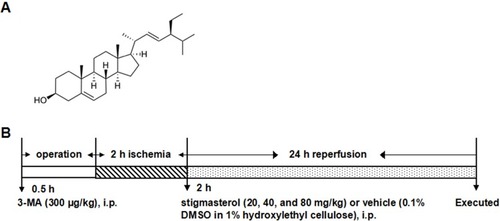
Figure 2 Effects of different doses of stigmasterol on infarct volumes (A, B), neurological grade (C), brain water content (D), and CBF (E) in cerebral ischemia/reperfusion injury. 3-MA was chosen as the positive control. n=6. ##p<0.01 vs sham group; *p<0.05, **p<0.01 vs ischemia/reperfusion group.
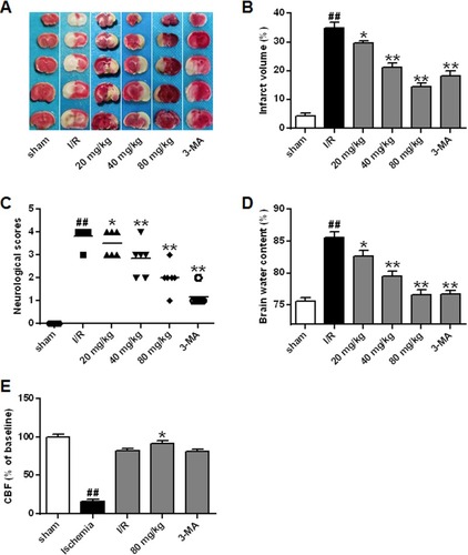
Figure 3 Effects of different doses of stigmasterol on histopathological alteration following cerebral ischemia/reperfusion injury (200×). After deep anesthetization with chloral hydrate, rat brain was immediately removed and immersed in 10% phosphate-buffered formalin for 12 hrs, then embedded in paraffin. Brain tissues of 5 μm were stained with HE and visualized. Arrows in the sham group, denoting the normal neurons; in ischemia/reperfusion group, the physiological abnormality; in stigmasterol-treated groups, the recovering neurons. The abnormal neurons were counted and expressed relatively to the sham group. n=6. ##p<0.01 vs sham group; *p<0.05, **p<0.01 vs ischemia/reperfusion group.
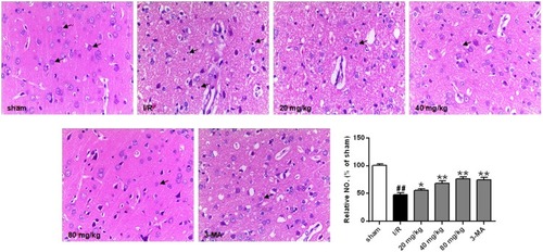
Figure 4 Effects of different doses of stigmasterol on the levels of nitric oxide (NO) (A), glutathione disulfide (GSSG) (B), malondialdehyde (MDA) (C), glutathione (GSH) (D) and the activities of SOD and its isoforms (E–G) and glutathione peroxidase (GSH-PX) (H) following cerebral ischemia/reperfusion injury. 3-MA was chosen as the positive control. n=6. ## p<0.01 vs sham group; *p<0.05, **p<0.01 vs ischemia/reperfusion group.
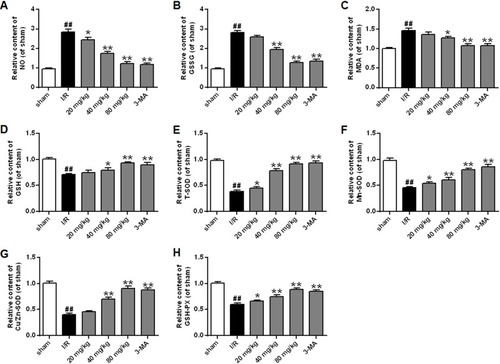
Figure 5 Effects of different doses of stigmasterol on the expression of beclin1 (A) and LC3 (B) of rats with cerebral ischemia/reperfusion injury. 3-MA was chosen as the positive control. n=6. ## p<0.01 vs sham group; *p<0.05, **p<0.01 vs ischemia/reperfusion group.
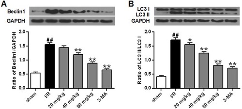
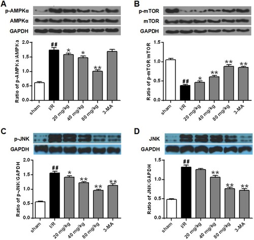
Figure 6 Effects of different doses of stigmasterol on the expression and phosphorylation of AMPKα (A), mTOR (B), and JNK (C, D) in rats with cerebral ischemia/reperfusion injury. 3-MA was chosen as the positive control. n=6. ## p<0.01 vs sham group; *p<0.05, **p<0.01 vs ischemia/reperfusion group.