Figures & data
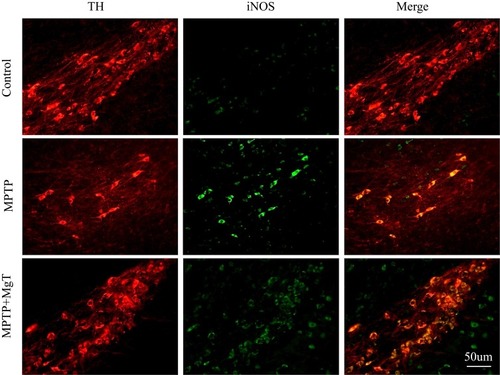
Figure 1 The changes of magnesium concentration in cerebrospinal fluid (CSF) and serum following treatment with MgSO4 or MgT on day 0, 21, and 28 in normal mice. *P<0.05, **P<0.01, compared with basal, #P<0.05, compared with MgSO4 group.
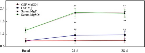
Figure 2 Open-field and rotarod tests. (A) Representative motor activity maps of mouse movement from each treatment group (day 28); (B) total distance; (C) average velocity and movement time; (D) rotarod time in 10, 12, 14, and 16 rpm/min, respectively. **P<0.01, compared with control. #P<0.05, compared with MPTP group.
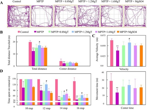
Figure 3 Immunohistochemical staining and Western blot analysis of TH. (A) Representative light micrographs of immuno-staining of TH in SN and striatum; (B) number of TH-positive cells in SNpc; (C) Western blot analysis of TH in SN and striatum. Upper row showed representative gel bands for TH protein, lower row showed β-actin signals from the same blot; (D) ration TH/β-actin. *P<0.05, **P<0.01, compared with control, #P<0.05, compared with MPTP group.
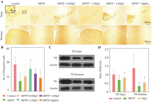
Figure 4 Immunohistochemical staining (A) and Western blot analysis of iNOS in SN; (B) representative light micrographs of immuno-staining of iNOS in SN were given in (A) quantitative analysis of iNOS-positive cells was presented in (B) **P<0.01, compared with control; #P<0.05, ##P<0.01, compared with MPTP group.
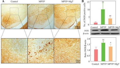
Figure 5 The results of immunofluorescence staining of iNOS/TH. Representative micrographs indicate most of the iNOS-positive cells were TH-positive DA neurons.