Figures & data
Table 1 Sequences of Primers Used in qRT-PCR
Figure 1 The miR-488-3p level was apparently reduced after OGD/R in vitro or I/R in vivo. (A) After OGD treatment in neuronal cells, mRNA level of miR-488-3p was diminished. (B) In mice undergoing transient MCAO/R, the miR-488-3p level was also diminished. (C) FISH assay was performed to observe the alteration of miR-488-3p in the hippocampus and cortex after I/R. Data were expressed as mean±SD. **P<0.01 (n = 6).
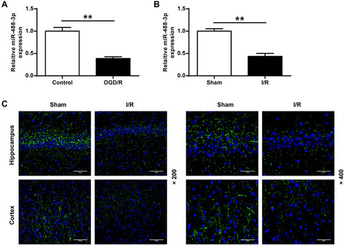
Figure 2 Up-regulation of miR-488-3p protected neurons against the injury induced by OGD/R in vitro. (A) The miR-488-3p expression level of neurons treated with miR-488-3p mimic or miR-488-3p inhibitor was valued by qRT-PCR. (B) MTT assay was applied to detect the cell viability. (C) The proportion of apoptotic cells was analyzed by Annexin V-FITC/PI staining. Data were expressed as mean±SD. *P<0.05, **P<0.01 (n=3).
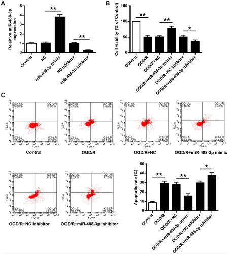
Figure 3 Up-regulated miR-488-3p attenuated ischemic brain injury in vivo. (A) After MCAO/R for 24 hours, miR-488-3p level was improved and diminished in brain treated with miR-488-3p mimic and miR-488-3p inhibitor, respectively. In mice with cerebral I/R, impact of miR-488-3p on neurobehavioral outcomes (B) and infarct volume (C) were detected. Data were expressed as mean±SD. *P<0.05, **P<0.01 (n=6).
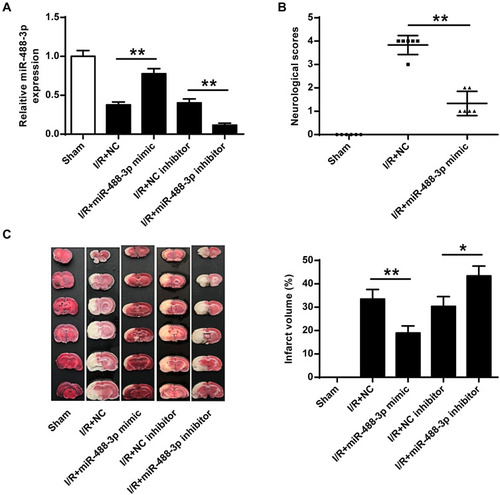
Figure 4 MiR-488-3p directly suppressed VPS4B. (A) Predicted interaction between miR-488-3p and VPS4B. (B) The vector map for the reporter clone. (C) After 48 hours transfection, the 3′-UTR reporter assay was conducted to confirm the luciferase expression. The luciferase activity in miR-488-3p mimic + VPS4B 3′-UTR WT group was apparently suppressed. (D) mRNA and (E) protein levels of VPS4B, after OGD/R for 24 hours, were measured in the miR-488-3p overexpressed/inhibited neurons. Data were expressed as mean±SD. **P<0.01 (n=3).
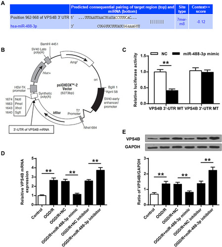
Figure 5 Down-regulated VPS4B expression suppressed neuronal cell death in vitro and ischemic brain injury in vivo. mRNA level of VPS4B (A) and protein level of VPS4B (B) dropped in neurons transfected with sh-VPS4B. (C) MTT assay was applied to analyze the cell viability. (D) Annexin V-FITC/PI staining was utilized to analyze the proportion of apoptotic cells. VPS4B mRNA level (E) and VPS4B protein level (F) was reduced in the I/R+ sh-VPS4B group, in comparison with the I/R+sh-NC group. In mice with cerebral I/R, effects of sh-VPS4B on neurobehavioral outcomes (G) and infarct volume (H) were analyzed. Data were expressed as mean±SD. *P<0.05, **P<0.01 (n=6).
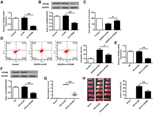
Figure 6 Up-regulated miR-488-3p attenuated ischemic brain injury by targeting VPS4B. (A) MTT assay was adopted to detect the cell viability. (B) Annexin V-FITC/PI staining was applied to assess the proportion of apoptotic cells. (C) TTC staining was utilized to evaluate the infarct volume in mice with cerebral I/R. Data were expressed as mean±SD. *P<0.05, **P<0.01 (n=6).
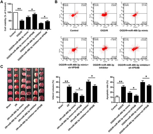
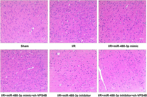
Figure 7 Impacts of miR-488-3p on cerebral morphological changes. Morphological detection by HE staining in groups of Sham, I/R, I/R + miR-488-3p mimic, I/R + miR-488-3p mimic + sh-VPS4B, I/R + miR-488-3p inhibitor, I/R +miR-488-3p inhibitor + sh-VPS4B (n=6).