Figures & data
Figure 1 A 13-year-old girl’s cAVMs removed via the transcranial neuroendoscopic approach. (A) CT scan showing subarachnoid hemorrhage and a small hematoma in the left posterior medial temporal gyrus. (B) CTA showing a small AVM in the temporal lobe. (C) DSA confirming a small AVM in the temporal lobe. (D) The intracranial pressure was high after opening the dura mater. (E) The hematoma was evacuated by neuroendoscopy. (F) Intracranial pressure decreased after the hematoma was removed. (G) The dilated and red-colored draining veins were discovered. (H) The nidus of the AVM was circumferentially dissected. (I) The draining vein became darker after the AVM was removed. (J) Postoperative CT scan showing that the hematoma had been completely removed. (K) Postoperative CTA showed that the AVM had disappeared. (L) Histopathologically confirmed AVMs.
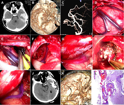
Figure 2 Preoperative and postoperative DSA images of the 13-year-old girl’s cAVMs. (A) Preoperative anteroposterior views show a small AVM supplied by a branch of the posterior cerebral artery. (B) Preoperative lateral views show that the vein was draining into the superior petrosal sinuses. (C) Preoperative views from anteroposterior 3D DSA showing the AVM structure. (D) Preoperative views from lateral 3D DSA showing the AVM structure. (E and F) Postoperative anteroposterior and lateral views showing that the AVM had disappeared. (G and H) Postoperative anteroposterior and lateral views from 3D DSA showing that the AVM had been completely excised.
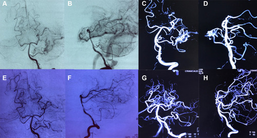
Figure 3 A 67-year-old man’s cAVMs removed via the transcranial neuroendoscopic approach. (A) CT scan at onset showing a hematoma in the right frontal lobe. (B) The hematoma was enlarged. (C) CTA showing a small AVM in the left frontal lobe. (D) A linear skin incision was made parallel to the Sylvain fissure. (E) Postoperative CT scan showing that the hematoma had been completely removed. (F) Postoperative image showing that the diameter of the bone flap was approximately 4 cm. (G) Postoperative CTA showing that the AVM had been completely removed. (H) Histopathologically confirmed arteriovenous malformations.
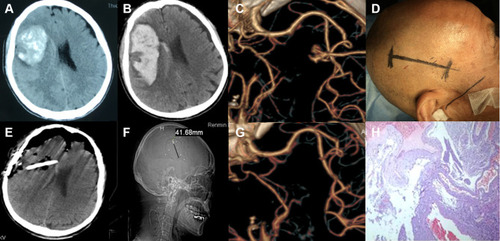
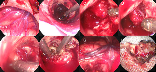
Figure 4 Surgical procedure of cAVM removal in the 67-year-old man via the transcranial neuroendoscopic approach. (A) After opening the dura mater, a red-colored draining vein was found. (B) The hematoma was evacuated under the visualization of the neuroendoscope. (C) The AVM nidus was located. (D) The feeding artery of the AVM was located. (E) The draining vein was identified. (F) The feeding artery was isolated, cauterized and cut. (G) The draining vein became darker after the AVM was removed. (H) The operative sample of the AVM.