Figures & data
Table 1 Clinical characteristics of the patients and distances between mappings by navigated transcranial magnetic stimulation and direct electrical cortical stimulation
Figure 1 (A) Mapping with DES after craniotomy with a bipolar-stimulation-system-like neuronavigation tool. (B) Neuronavigation system image corresponding to this point in DES mapping in a three-dimensional, axial, sagittal, and coronal slice.
Abbreviation: DES, direct electrical cortical stimulation.
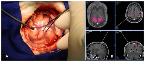
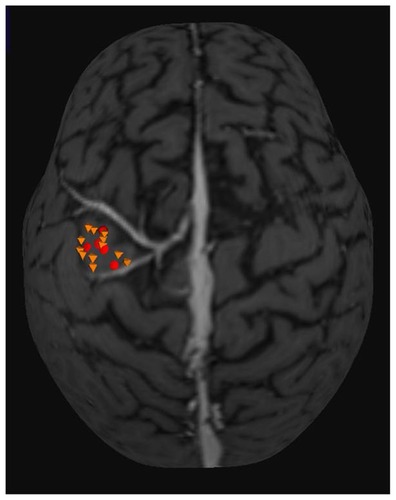
Figure 2 Three-dimensional MRI used in neuronavigation system. Yellow octahedrons are points with motor response recorded in the TMS mapping. Red circles are the points with the motor response in DES mapping.
Note: The mean distance between the two mappings found was 4.16 mm.
Abbreviations: DES, direct electrical cortical stimulation; MRI, magnetic resonance imaging; TMS, transcranial magnetic stimulation.
Abbreviations: DES, direct electrical cortical stimulation; MRI, magnetic resonance imaging; TMS, transcranial magnetic stimulation.