Figures & data
Table 1 A summary of the findings
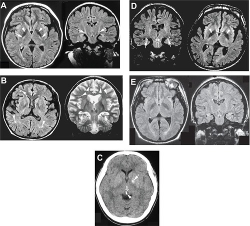
Figure 1 (A) FLAIR sequence of brain MRI at the age of 10 years (Case 1). The patient showed marked areas of low signal intensity in the pallidum (arrowhead) and areas of slightly high signal intensity in the pyramidal tract (arrow). (B) FLAIR and T2-weighted sequences of MRI at the age of 30 years (Case 2). The patient showed hippocampal atrophy (arrow) and diffuse mild ischemic changes in the cerebral white matter (arrowhead). (C) CT scan at the age of 28 years (Case 3). The patient showed calcification of the pallidum (arrow), pineal body (arrowhead), and habenular commissure (open arrow). (D) FLAIR sequences of MRI at the age of 26 years (Case 4). The patient showed hippocampal atrophy (arrow), marked areas of low signal intensity in the pallidum (arrowhead), and areas of increased signal intensity in the decussation of the superior cerebellar peduncles (open arrow). (E) FLAIR sequences of MRI at the age of 18 years (Case 5). The patient showed hippocampal atrophy.