Figures & data
Table 1 Differences in semiology and timing between PNES and epileptic seizures
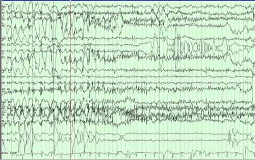
Figure 1 The PT transitions into a typical PNES.
Notes: Immediately prior to it, the EEG background shows a normal waking rhythm. The EEG is then obscured by movement and muscle artifact that is most prominent in the posterior regions.
Abbreviations: PT, patient; PNES, psychogenic nonepileptic seizure; EEG, electroencephalogram.
Abbreviations: PT, patient; PNES, psychogenic nonepileptic seizure; EEG, electroencephalogram.
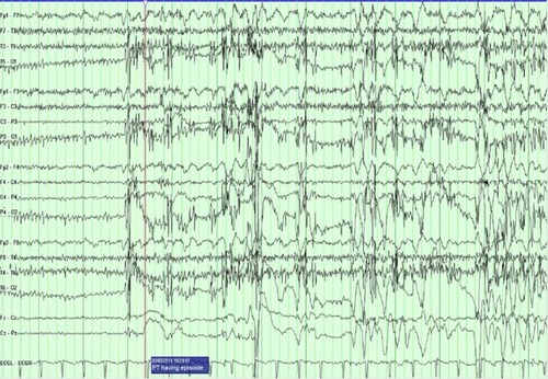
Figure 2 The patient is in the middle of a typical PNES.
Notes: The EEG background is obscured by movement and muscle artifact that is most prominent in the posterior regions. There is an embedded 1-second period when the movement stops and normal background is seen before the background is again obscured by movement and muscle artifact.
Abbreviations: PNES, psychogenic nonepileptic seizure; EEG, electroencephalogram.
Abbreviations: PNES, psychogenic nonepileptic seizure; EEG, electroencephalogram.
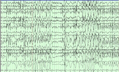
Figure 3 The patient transitions out of a typical PNES.
Notes: Initially, the slide shows EEG background obscured by movement and muscle artifact that is most prominent in the posterior regions. Immediately after it, the EEG background returns to a normal waking background.
Abbreviations: PNES, psychogenic nonepileptic seizure; EEG, electroencephalogram.
Abbreviations: PNES, psychogenic nonepileptic seizure; EEG, electroencephalogram.