Figures & data
Figure 1 Bar graph showing the result of multiplex ligation-dependent probe amplification (MLPA) analysis using the SALSA MLPA P081 and P082 NF1 kits (MRC-Holland, Amsterdam, the Netherlands). Relative amounts of probe-amplified products were compared with reference samples and data analysis was performed using the Coffalyser 9.4 package (MRC-Holland). Values under a threshold of 0.7 and over a threshold of 1.3 for multiple adjacent probes indicate the presence of a deletion or duplication, respectively. The arrows highlight the exons 12 (10a) and 13 (10b) deleted in this patient.
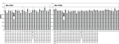
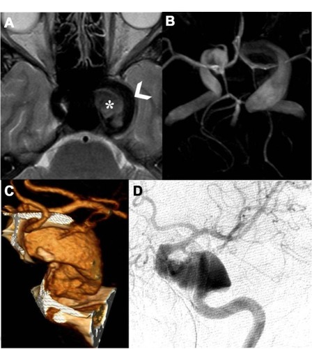
Figure 2 Magnetic resonance (MR) axial T2-weighted cerebral study shows flow-void signal of patent intracavernous side carotid siphon (arrowhead), and, medially, intravascular characteristic signal modification to refer breakdown hemoglobin products of thrombus (asterisk) (A); time-of-flight MR angiography (TOF-MRA), using a maximum-intensity projection (MIP) algorithm with 3-D MIP reconstruction, detailed two intracranial fusiform aneurysms, one at the left internal carotid artery (ICA) as a giant fusiform intracavernous aneurysm (larger than 25 mm in diameter), and the second as a contralateral smaller aneurysm (12 mm larger) (B); angio-computed tomography (A-CT) shaded surface display reconstruction (SSD) study shows magnified detail of left partially thrombosed fusiform giant aneurysm (C); left selective ICA digital angiography anteroposterior view confirms the partially thrombosed giant intracavernous aneurysm (D).