Figures & data
Figure 1 Structures of GABA and VGB.
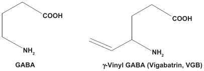
Figure 2 Schematic demonstrating a common target of action shared by many forms of therapy. Treatment with adrenocorticotropic hormone stimulates the adrenal production of tetrahydrodeoxycorticosterone which can activate extrasynaptic gamma aminobutyric acid receptors which mediate tonic inhibition. Increased ambient gamma aminobutyric acid produced by vigabatrin treatment may have a similar effect, as also the treatment with ganaxalone a neurosteroid investigational drug. The α4βxδ subunit containing extrasynaptic receptors are readily activated by modest levels of ambient gamma aminobutyric acid to produce a “lasting” current to sustain tonic activation. Adapted with permission from Auvin S, Sankar R. Antiinflammatory treatments for seizure syndromes and epilepsy. In: Rho JM, Sankar R, Stafstrom CE, editors. Epilepsy: Mechanisms, Models, and Translational Perspectives. New York, NY: CRC Press/Taylor and Francis; 2010.
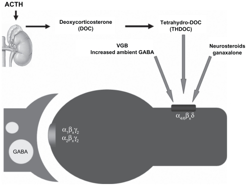
Table 1 Clinical studies of vigabatrin in the treatment of infantile spasms
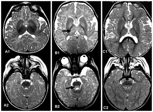
Figure 3 A1 and A2) show axial T2 weighted images of a normal baby aged five months. B1 and B2) show a five-month-old patient who had received vigabatrin for eight weeks. The magnetic resonance image was obtained due to abnormal eye movement. Magnetic resonance imaging shows high T2 weighted intensity in the thalamus and globus pallidus (arrow in B1) and tegmental portion of the pons (arrow in B2). Vigabatrin was discontinued. C1 and C2) Follow-up magnetic resonance imaging of the same patient three months later shows normal signal in the thalamus and pontine tegmentum.