Figures & data
Figure 1 Computed tomography perfusion with four regions of interest selected in each cerebral hemisphere.
Note: Three superficial regions and one positioned in the basal ganglia.
Abbreviations: R, right; P, posterior.
Abbreviations: R, right; P, posterior.
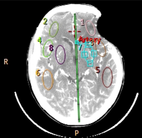
Table 2 Summary of case series reported
Figure 2 Cranial computed tomography before performing cranioplasty.
Note: We observed a significant depression of the skin flap with erasing wrinkles and superficial cortical compression.
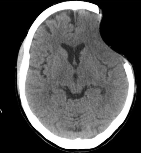
Table 1 Neuropsychological assessment before and 6 months after cranioplasty
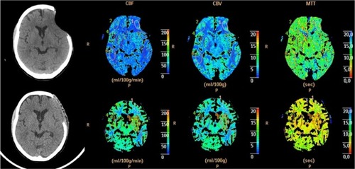
Figure 3 Perfusion computed tomography before (top) and after (bottom) surgery showing the changes in the three parameters: CBF, CBV, and MTT.
Note: In this image we can also see a restructuring of the cortical mantle of the left frontal region.
Abbreviations: CBF, cerebral blood flow; CBV, cerebral blood volume; MTT, mean transit time.
Abbreviations: CBF, cerebral blood flow; CBV, cerebral blood volume; MTT, mean transit time.