
Figures & data
Figure 1 CT of cranial bones.
Abbreviation: CT, computed tomography.
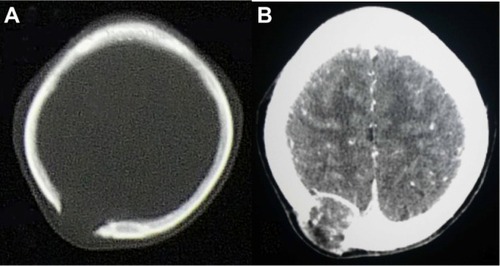
Figure 2 MRI of cranial bones.
Abbreviation: MRI, magnetic resonance imaging.
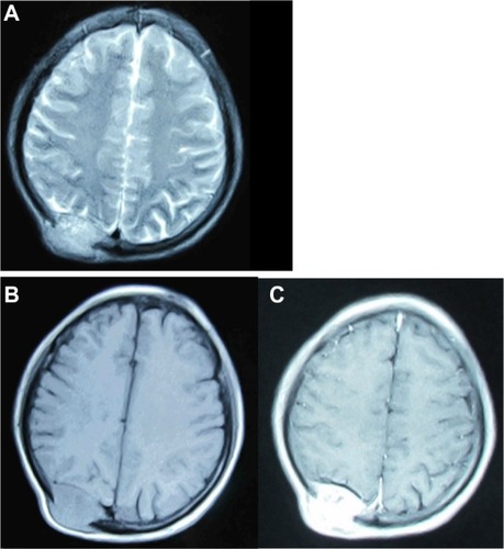
Figure 3 Abdominal B ultrasound showed a large mass in the right lobe of the liver.
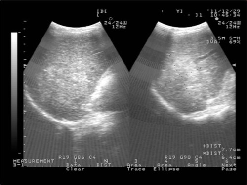
Figure 4 Contrast-enhanced computed tomography of abdomen showed a huge enhanced carcinoma in the liver.
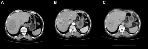
Figure 5 Computed tomography (CT) of breast showed no lung metastases.
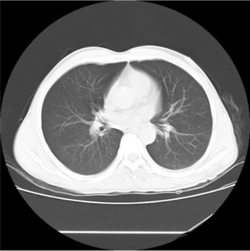
Figure 6 Single-photon emission computed tomography of total skeletal bones showed no other metastases.
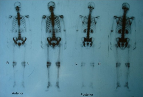
Table 1 Laboratory data on admission
Figure 7 The histopathological characteristics of carcinoma and the immunohistochemical finding.
Abbreviations: HE, hematoxylin-eosin staining; AFP, alpha fetal protein; CK, creatine kinase; CD, cluster of differentiation or leukocyte differentiation antigen; Ki67, nuclear-associated antigen Ki67.
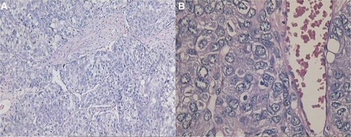
Table 2 Summary of reported cases in the literature with skull metastases from HCC (n=59)Table Footnote*
Table 3 Summary of reported cases in the literature with skull metastases as the first symptom from HCC (n=33)