Figures & data
Figure 1 Brain magnetic resonance imaging (MRI) findings.
Notes: (A and B) MRI (diffusion-weighted image) on admission; (C and D) MRI (diffusion-weighted image) on hospital day 22; (E and F) MRI (diffusion-weighted image) on hospital day 36; and (G and H) MRI (diffusion-weighted image) on hospital day 66. Figures in the top set (A, C, E, and G) are images at the cerebral level of corona radiata. Figures in the bottom set (B, D, F, and H) are images at the cerebral level of lenticular nuclei. “R” indicates the right side of the patient’s brain.
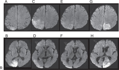
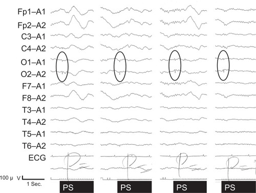
Figure 2 Electroencephalographic findings on hospital day 57.
Note: Positive sharp waves were evoked by photic stimulation in bilateral occipital leads as shown in the ovals.
Abbreviations: ECG, electrocardiogram; PS, photic stimulation; sec, second.
Abbreviations: ECG, electrocardiogram; PS, photic stimulation; sec, second.