Figures & data
Figure 1 Axial T2-weighted magnetic resonance imaging (MRI) of the body of the lateral ventricle showing a single infarction in the left corona radiata.
Notes: (A) The arrow points to the major pathological feature. (B) Axial T2-weighted MRI of the basal ganglia showing periventricular hyper-intensity and subcortical white matter hyperintensities in both hemispheres, without major brain infarction.
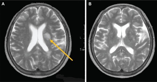
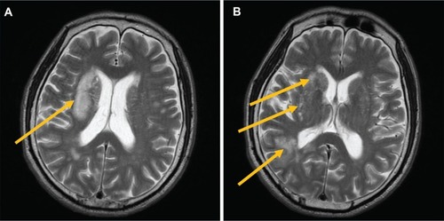
Figure 2 Axial T2-weighted magnetic resonance imaging (MRI) of the body of the lateral ventricle showing infarction of the right corona radiata, without major brain infarction in the left hemisphere.
Notes: (A) Axial T2-weighted MRI of the basal ganglia showing infarction of the caudate nucleus, lentiform nucleus, and right posterior temporal lobe. (B) Subcortical white matter hyperintensities in both hemispheres are present. Arrows point to the major pathological features.