Figures & data
Figure 1 X-ray of the cervical spine anterior–posterior (A) and lateral (B) views showed a mild narrowing of intervertebral disk spaces at almost every level of the cervical spine and osteoarthrosis of several apophyseal joints.
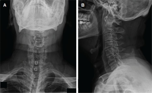
Figure 2 Cervical spine magnetic resonance imaging sagittal (A) and axial views showed herniation of the intervertebral disk in C3/4 (B1), C4/5 (B2), and C5/6 (B3) shown by arrows in (A), with a compression of the ventral surface of the spinal cord.
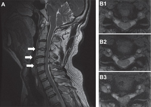
Figure 3 Axial view of brain computed tomography without contrast.
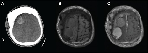
Figure 4 A–D Histologic findings of atypical meningiomas (World Health Organization) grade II.
Abbreviations: HPF, high power fields; EMA, epithelial membrane antigen.
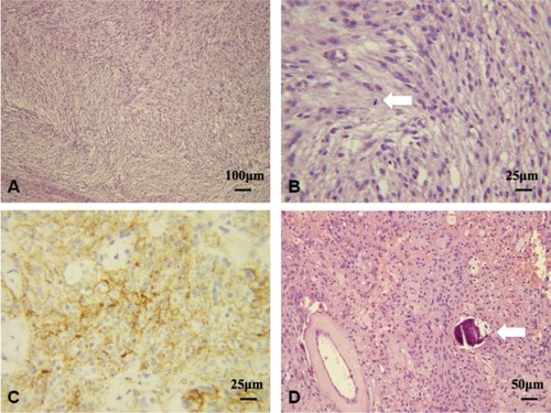
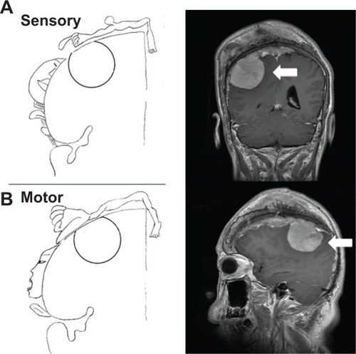
Figure 5 Depending on the location of the patient’s meningioma (arrows), the weakness, numbness, and paresthesia of the upper-left limb were compatible with the cortical homunculus, which compressed to the primary somatosensory area (A) and the primary motor cortex (B).