Figures & data
Figure 1 The ictal EEG in case 1.
Notes: (A) Case 1, third session. There was no clear progression to the slow-wave phase. The amplitudes of slow waves were low. The peak amplitude was 160 μV (45 seconds). The EMG endpoint was 44 seconds, and the EEG endpoint was obscure. (B) Case 1, fourth session. The onset of the slow-wave phase was distinguishable. High-amplitude slow waves were observed from 47 to 58 seconds. The peak amplitude was 360 μV (51 seconds). The EMG/EEG endpoints were 54/78 seconds. Post-ictal suppression was achieved, but the transition to flat was gradual. (C) Case 1, sixth session. The latency to slow waves was relatively short. High amplitude slow waves were regularly observed. Peak amplitude was 440 μV (51 seconds). EMG/EEG endpoints were 48/63 seconds. Post-ictal suppression was achieved, but the transition to flat was still gradual.
Abbreviations: EMG, electromyographic; EEG, electroencephalographic.
Abbreviations: EMG, electromyographic; EEG, electroencephalographic.
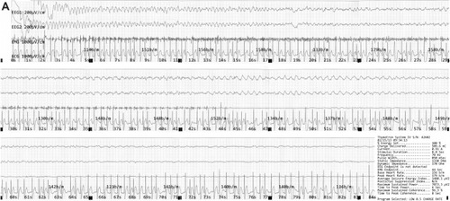
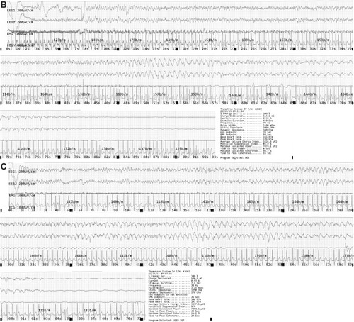
Figure 2 The ictal EEG in case 2.
Notes: (A) Case 2, fifth session. Ictal slow waves were not observed. (B) Case 2, sixth session. Irregular slow waves were observed. The peak amplitude was 320 μV (36 seconds). The EMG/EEG endpoints were 94/114 seconds. Seizure termination was clear, but suppression was poor. (C) Case 2, ninth session. Irregular slow waves were observed again after the failure in the eighth session. The peak amplitude was 230 μV (42 seconds). The EMG/EEG endpoints were 48/53 seconds. Seizure termination was distinguishable, but suppression appeared poor. The post-seizure monitoring time was probably insufficient.
Abbreviations: EMG, electromyographic; EEG, electroencephalographic.
Abbreviations: EMG, electromyographic; EEG, electroencephalographic.