Figures & data
Table 1 Classification of stroke
Figure 1 Risk factors and dementia.
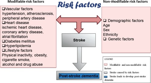
Figure 2 Clinical evaluation.
Abbreviations: CT, computed tomography; ECG, electrocardiography; EEG, electroencephalography; MRI, magnetic resonance imaging.
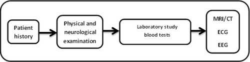
Figure 3 Core and penumbra after stroke.
Note: Reprinted from Journal of Radiology Nursing, 30(3), Summers D, Malloy R, CT and MR imaging in the acute ischemic stroke patient: a nursing perspective,104–115, Copyright 2011, with permission from Elsevier.Citation56
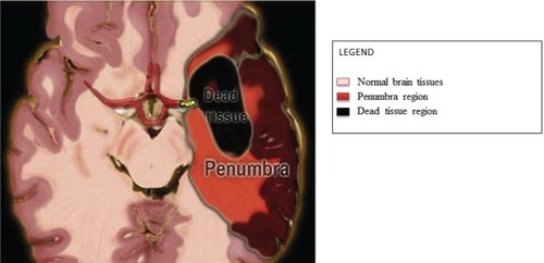
Table 2 Stroke outcome due to vessel infarction
Figure 4 Block diagram of vascular cognitive impairment spectrum.
Abbreviations: AD, Alzheimer’s disease; CVD, cerebral vascular disease; MCI, mild cognitive impairment; PSD, post-stroke dementia; VaD, vascular dementia; VCI, vascular cognitive impairment.
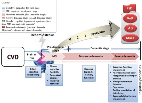
Table 3 Types of memory
Table 4 Brain memory loss causes
Table 5 Memory classification
Table 6 Neuropsychological assessment characteristics