Figures & data
Figure 1 Axial computed tomography study of the brain.
Notes: Minute cortical and subcortical calcifications located in bilateral frontal lobe.
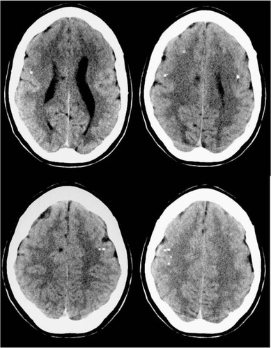
Figure 2 Axial Spin-Echo T2-weighted (A, C) and T1-weighted (B, D) magnetic resonance imaging study of the brain.
Notes: Developmental abnormalities (polymicrogyria) in the frontal opercular cortex bilaterally, more evident on the left side.
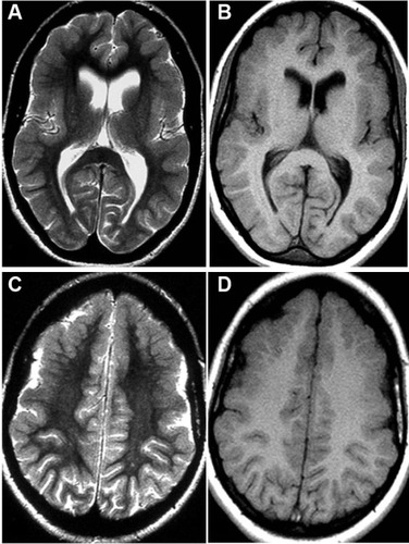
Figure 3 Spin-Echo T2-weighted sagittal projections on magnetic resonance imaging study of the brain.
Notes: A “cobblestone” appearance is evident in the bilateral frontal opercular region.
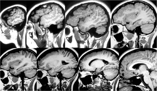
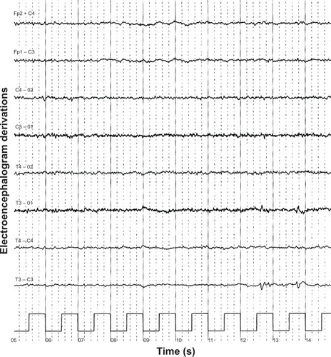
Figure 4 Electroencephalogram showing diffusely deregulated cerebral electrical activity together with ubiquitous fast paroxysms, prevalent in the left temporal area.