
Figures & data
Table 1 Demographic and clinical findings of OCD patients (n=39) and healthy participants (n=30)
Table 2 Clinical findings before and after behavior therapy in the OCD groups (n=39)
Figure 1 (A) Smaller regional GM volumes were observed for the right orbitofrontal cortex and left cingulate cortex in nonresponder OCD patients (n=15), compared with responder OCD patients (n=24). (B) Smaller regional WM volumes were observed for the left cingulate bundle in nonresponder OCD patients (n=15), compared with responder OCD patients (n=24).
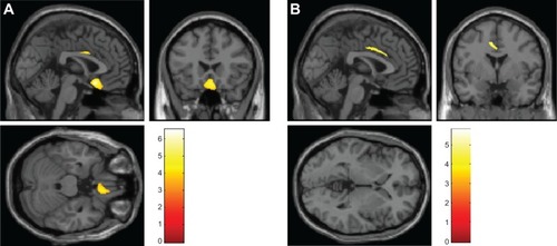
Table 3 Areas with smaller regional GM and WM volumes in nonresponder OCD patients (n=15) than in responder OCD patients (n=24)
Table 4 Areas with smaller regional GM and WM volumes in nonresponder OCD patients (n=15) than in healthy participants (n=30)
Table 5 Areas with smaller regional GM and WM volumes in responder OCD patients (n=24) than in healthy participants (n=30)