
Figures & data
Table 1 Demographic data
Figure 1 Task design.
Notes: Each block lasted 30 seconds and consisted of ten trials. Each trial consisted of a 2 second visual stimulus (BM, SM, or ST) and a 1 second interval (white cross). Each session consisted of 12 seconds that were discarded prior to analysis, followed by eight blocks. Each experiment involved three sessions.
Abbreviations: BM, biological motion; s, seconds; SM, scrambled motion; ST, static condition.
Abbreviations: BM, biological motion; s, seconds; SM, scrambled motion; ST, static condition.
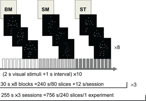
Figure 2 Brain regions significantly activated in the BM–ST (A, B) and SM–ST (C, D) conditions have been superimposed onto the SPM templates.
Notes: In both groups (normal and schizophrenia), the activated clusters covered the bilateral hMT+ and bilateral frontal regions in both the BM–ST and SM–ST conditions. Furthermore, in the SM–ST condition, the bilateral superior parietal lobules were activated in both the groups. A one-sample t-test, q(FDR)<0.05, and an extended threshold of ten contiguous voxels were employed. No significant activation was observed after subtraction of the results of schizophrenia group from those of normal control group in both the BM–ST and SM–ST conditions. A two-sample t-test, P<0.0005 (uncorrected) and an extended threshold of ten contiguous voxels were employed.
Abbreviations: BM, biological motion; FDR, false discovery rate; hMT+, motion-sensitive area; SM, scrambled motion; SPM, statistical parametric mapping; ST, static condition.
Abbreviations: BM, biological motion; FDR, false discovery rate; hMT+, motion-sensitive area; SM, scrambled motion; SPM, statistical parametric mapping; ST, static condition.
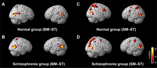
Figure 3 Brain regions significantly activated in the BM–SM conditions have been superimposed onto the SPM templates.
Notes: (A) Brain regions that were significantly activated in the normal group. The activated clusters covered the bilateral STS, bilateral medial frontal cortex, left precuneus, and middle cingulate. A one-sample t-test, q(FDR)<0.05, and an extended threshold of ten contiguous voxels were employed. (B) Brain regions that were significantly activated in the schizophrenia group. The activated clusters included bilateral STS only. A one-sample t-test, q(FDR)<0.05, and an extended threshold of ten contiguous voxels were employed. (C) Brain regions that were more activated in the normal group than in the schizophrenia group. The activated cluster included the left medial frontal gyrus, supramarginal gyrus, precuneus, cuneus, and middle cingulate. A two-sample t-test, P<0.0005 (uncorrected) and an extended threshold of ten contiguous voxels were employed. Z; z-value of each transverse slice.
Abbreviations: BM, biological motion; FDR, false discovery rate; hMT+, motion-sensitive area; SM, scrambled motion; SPM, statistical parametric mapping; STS, superior temporal sulcus.
Abbreviations: BM, biological motion; FDR, false discovery rate; hMT+, motion-sensitive area; SM, scrambled motion; SPM, statistical parametric mapping; STS, superior temporal sulcus.
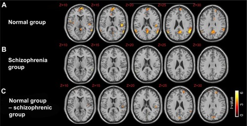
Table 2 Significantly activated clusters for the BM–ST contrast
Table 3 Significantly activated clusters for the SM–ST contrast
Table 4 Significantly activated clusters for the BM–SM contrast