
Figures & data
Table 1 Demographic characteristics according to the type of intracranial aneurysm
Figure 1 Relative prevalence of risk factors by age. (A) In patients with MirAn. (B) In nMirAn patients.
Note: Smoking refers to cigarette smoking.
Abbreviations: HBP, high blood pressure; MirAn, mirror-like intracranial aneurysms; nMirAn, non-mirror-like multiple aneurysms; NRF, no known extrinsic risk factors.
Abbreviations: HBP, high blood pressure; MirAn, mirror-like intracranial aneurysms; nMirAn, non-mirror-like multiple aneurysms; NRF, no known extrinsic risk factors.
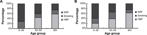
Table 2 Hunt and Hess grade and aneurysm sites in patients with SAH
Figure 2 DSA images of both the left and right internal carotid arteries before clipping. DSA suggests that the aneurysms arise from the branches of the bilateral middle cerebral artery (indicated by arrow).
Notes: (A) Right internal carotid artery (normal position); (B) right internal carotid artery (lateral position); (C) left internal carotid artery (normal position); (D) left internal carotid artery (lateral position).
Abbreviation: DSA, digital subtraction angiography.
Abbreviation: DSA, digital subtraction angiography.
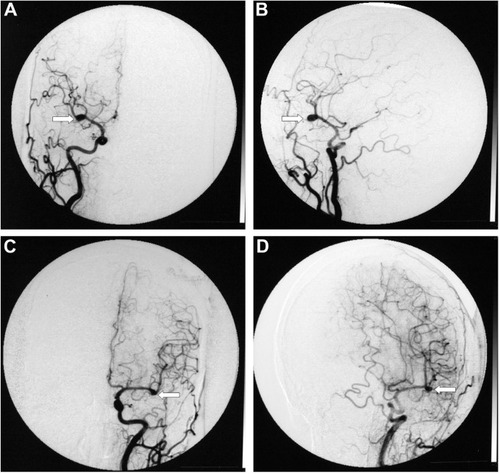
Figure 3 DSA images of the left and right internal carotid arteries after clipping. Both aneurysms from the bilateral middle cerebral artery branches are completely occluded (normal position, indicated by the arrow).
Notes: (A) Left aneurysms; (B) right aneurysms.
Abbreviation: DSA, digital subtraction angiography.
Abbreviation: DSA, digital subtraction angiography.
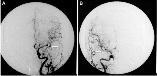
Table 3 Treatment and follow-up for the patients with MirAn