Figures & data
Figure 1 Transverse T1-weighted magnetic resonance images of the brain.
Notes: (A–C) Diffuse atrophy of the brain, including the cerebrum, cerebellum, and hippocampus was found, accompanied by dilatation of ventricles, subarachnoid space, and sulci, with an Evans’ index of 0.25. (D–F) Eight months after surgical removal of cortisol-secreting adrenocortical adenoma, brain atrophy recovered. Dilatation of ventricles, subarachnoid space, and sulci was also resolved, with an Evans’ index of 0.22.
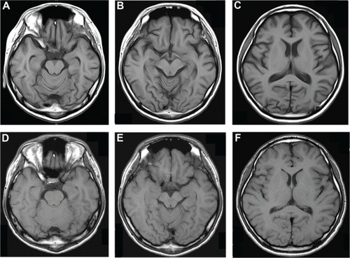
Figure 2 Radiological findings.
Notes: (A) Plain abdominal computed tomography showed a tumor of 2.7 cm diameter with 35 Hounsfield units (arrow) in the left adrenal gland. (B) 131I-adosterol scan (posterior view) demonstrated radioisotope accumulation in accordance with the left adrenal mass (arrow), while no uptake was detected on the opposite side.
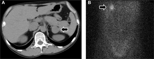
Table 1 Laboratory findings in August 2011
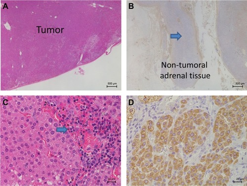
Figure 3 Histological findings of the resected left adrenal gland.
Notes: (A) Tissue sections stained with hematoxylin and eosin (HE) (20×) demonstrated an apparently marginated encapsulated tumor without any capsular or blood vessel invasion. (B) The zona reticularis and fasciculata of the attached non-tumoral adrenal tissue was atrophic (arrow) and showed little expression of dehydroepiandrosterone sulfate (20×). (C) The tumor consisted mainly of compact cells and partially of clear cells (HE, 400×). A slight infiltration of lymphocytes (arrow) was observed in the tumor. (D) Most of the tumor cells indicated positive immunostaining for 17α-hydoroxylase (400×).