Figures & data
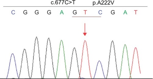
Figure 1 (A) T2-weighted sagittal magnetic resonance image of the cervical and thoracic spinal cord without contrast medium showing a hyperintense lesion with swelling at C7 to T3 (arrow). (B) A follow-up spinal magnetic resonance image 3 months later showed almost complete disappearance of the original lesions.
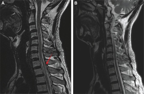
Figure 2 The patient has a homozygous MTHFR gene mutation c.677C>T (p.A222V).
Note: The position is indicated with an arrow.
Abbreviation: MTHFR, methylenetetrahydrofolate reductase.
Abbreviation: MTHFR, methylenetetrahydrofolate reductase.