Figures & data
Table 1 Effects of berberine on pilocarpine-induced convulsions in rats
Figure 1 Effects of berberine (Ber) on catalase (A), glutathione level (B), lipid peroxidation level (C), and superoxide dismutase (D) activities in hippocampus 24 hours after Pilo-induced status epilepticus in rats (n=4–6 in each group). Results are expressed as means ± standard error of mean.
Abbreviations: MDA, malondialdehyde; Pilo, pilocarpine; Ber 25, berberine 25 mg/kg; Ber 50, berberine 50 mg/kg; Ber 100, berberine 100 mg/kg.
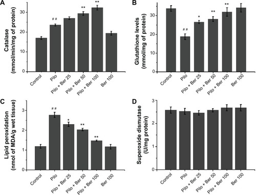
Figure 2 Effects of berberine (Ber) on latency to escape in Morris water-maze test 14 days after pilocarpine (Pilo)-induced status epilepticus (SE) in rats. Two-way repeat-measure analysis of variance revealed that Ber (50 and 100 mg/kg) significantly decreased escape latency in SE rats compared to saline-treated SE rats (P<0.05 and P<0.01, respectively; n=5 in each group). Results are expressed as means ± standard error of mean.
Abbreviations: Ber 25, berberine 25 mg/kg; Ber 50, berberine 50 mg/kg; Ber 100, berberine 100 mg/kg; SE, status epilepticus.
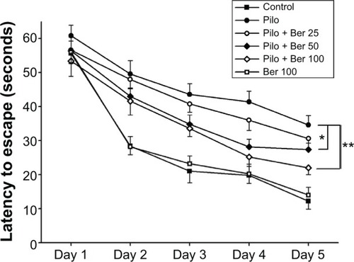
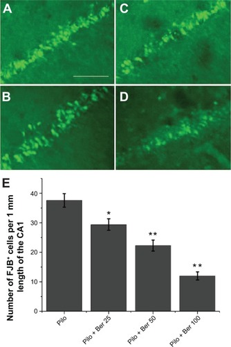
Figure 3 Effects of berberine (Ber) on number of fluoro-jade B (FJB)-positive cells in hippocampal CA1 region 20 days after pilocarpine (Pilo)-induced status epilepticus (SE) in rats (n=5 in each group). The non-SE control rats and Ber 100 mg/kg alone-treated rats had no FJB-positive cells in hippocampal CA1 (data not shown). FJB-positive cells were abundant in the CA1 of saline-treated SE rats (A). There were significantly fewer FJB-positive cells in the CA1 in the Pilo + Ber 50 group (B), Pilo + Ber 25 group (C), and Pilo + Ber 100 group (D) compared to saline-treated SE rats. Quantitative analysis of FJB-positive cells demonstrated that Ber reduced FJB-positive cells in CA1 (E).
Abbreviations: Ber 25, berberine 25 mg/kg; Ber 50, berberine 50 mg/kg; Ber 100, berberine 100 mg/kg.