Figures & data
Figure 1 Preoperative and postoperative findings by MRI and CT.
Notes: (A) Preoperative MRI scan showing the tumor located in the left part of the ventral medulla oblongata. (B) Postoperative day 1 CT scan showing no apparent ischemic infarct area. (C) Postoperative day 3 CT scan showing an ischemic infarction at the bilateral internal carotid arteries and the posterior cerebral artery. (D) Postoperative day 3 MRA showing stenosis in the siphon segment of the bilateral internal carotid arteries and distal segment of basilar artery, and the slender distal blood vessel. Cerebral artery showed a slender, segmental stricture. (E) Postoperative CT scan showing the progressively enlarged ischemic infarction area. (F) Postoperative CT scan showing decompressive craniectomy.
Abbreviations: CT, computed tomography; MRA, magnetic resonance angiography; MRI, magnetic resonance imaging.
Abbreviations: CT, computed tomography; MRA, magnetic resonance angiography; MRI, magnetic resonance imaging.
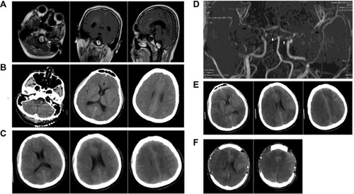
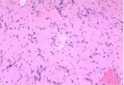
Figure 2 Photomicrograph of the tumor specimen.
Note: Pathological examination was consistent with a schwannoma.