Figures & data
Table 1 Diagnostic criteria for CLH
Figure 1 MRI sections showing multiple demyelinating brain lesions (arrows).
Notes: MRI scans of our presented case, showing multiple focal demyelinating changes, up to 11 mm in diameter: hyperintense in T2w (sagittal section, left image) and FLAIR (axial section, middle image), isointense in T2w (coronal section, right image) localized in the supratentorial frontotemporoparietal white matter, supra- and periventricular corners of the lateral chambers on both sides, in the centrum semiovale, corpus callosum, basal ganglia, and in the left perithalamic region. Also, lesions were found dorsolaterally in the right side of pons, and in the left middle cerebellar peduncle.
Abbreviations: FLAIR, fluid attenuated inversion recovery; MRI, magnetic resonance imaging; T2w, T2-weighted.
Abbreviations: FLAIR, fluid attenuated inversion recovery; MRI, magnetic resonance imaging; T2w, T2-weighted.
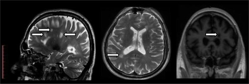
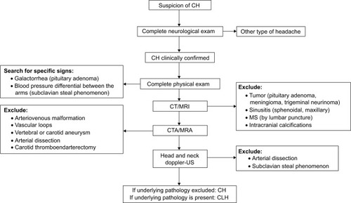
Figure 2 Protocol for investigation of CH patients.
Abbreviations: CH, cluster headache; CLH, cluster-like headache; CT, computed tomography; CTA, computed tomography angiography; MRI, magnetic resonance imaging; MRA, magnetic resonance angiography; MS, multiple sclerosis; US, ultrasound.