Figures & data
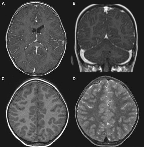
Figure 1 Brain MRI of a 3-year-old girl with anti-N-methyl-d-aspartate receptor encephalitis.
Notes: T1-weighted with gadolinium-enhanced MRI revealed (A) prominence of leptomeningeal enhancement at bilateral frontoparietal areas (axial view) and (B) nodular enhancement along the tentorium edge (coronal view). (C) T1-weighted (axial view) and (D) T2-weighted (axial view) MRI showing tiny white-matter lesions over bilateral frontal lobes.
Abbreviation: MRI, magnetic resonance imaging.
Abbreviation: MRI, magnetic resonance imaging.