172
Views
26
CrossRef citations to date
0
Altmetric
Original Research

Connectivity pattern differences bilaterally in the cerebellum posterior lobe in healthy subjects after normal sleep and sleep deprivation: a resting-state functional MRI study

, , , , , & show all
Pages 1279-1289 | Published online: 26 May 2015

Figures & data

Figure 1 Altered resting-state functional connectivity (rsFC) areas of the cerebellum posterior lobe (CPL) in the normal sleep group.

Notes: The rsFC areas of the left CPL were seen in the left precuneus, right inferior parietal lobule, bilaterally in the CPL, bilaterally in the superior frontal gyrus, and bilaterally in the middle frontal gyrus (A), while the rsFC areas of the right CPL were seen in the left middle frontal gyrus, left anterior cingulate, left angular gyrus, right posterior cingulate, right inferior parietal lobule, right cerebellum anterior lobe, right CPL, bilaterally in the superior frontal gyrus, bilaterally in the middle temporal gyrus, bilaterally in the inferior temporal gyrus, and supramarginal gyrus (B).
Figure 1 Altered resting-state functional connectivity (rsFC) areas of the cerebellum posterior lobe (CPL) in the normal sleep group.

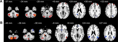
Table 1 Altered resting-state functional connectivity areas of the left cerebellum posterior lobe after normal sleep

Table 2 Altered resting-state functional connectivity areas of the right cerebellum posterior lobe after normal sleep

Figure 2 Altered resting-state functional connectivity (rsFC) areas of the cerebellum posterior lobe (CPL) in the sleep deprivation group.

Notes: The rsFC areas of the left CPL were seen in the left precentral gyrus, left angular gyrus, left inferior frontal gyrus, left superior parietal lobule, left inferior parietal lobule, left postcentral gyrus, left precuneus, left posterior cingulate gyrus, right middle frontal gyrus, and bilaterally in the CPL (A), while the rsFC areas of the right CPL were seen in the left superior frontal gyrus, left middle frontal gyrus, left paracentral lobule, left superior temporal gyrus, left middle temporal gyrus, bilaterally in the cerebellum anterior lobe, and bilaterally in the CPL (B).
Figure 2 Altered resting-state functional connectivity (rsFC) areas of the cerebellum posterior lobe (CPL) in the sleep deprivation group.

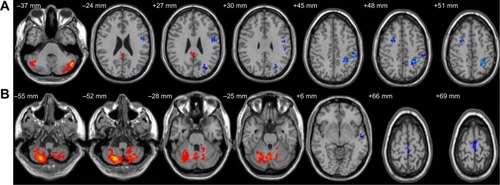
Table 3 Altered resting-state functional connectivity areas of the left cerebellum posterior lobe after sleep deprivation

Table 4 Altered resting-state functional connectivity areas of the right cerebellum posterior lobe after sleep deprivation

Figure 3 Altered resting-state functional connectivity (rsFC) areas of the cerebellum posterior lobe (CPL) in the sleep deprivation group compared with in the normal sleep group.

Notes: The rsFC areas of the left CPL were seen in the left middle frontal gyrus, right fusiform temporal gyrus, right cingulate gyrus, right thalamus, bilaterally in the superior frontal gyrus, bilaterally in the inferior frontal gyrus, bilaterally in the precuneus (A), while the rsFC areas of the right CPL were seen in the left precentral gyrus, left inferior frontal gyrus, right superior temporal gyrus, bilaterally in the superior frontal gyrus, and middle frontal gyrus (B).
Figure 3 Altered resting-state functional connectivity (rsFC) areas of the cerebellum posterior lobe (CPL) in the sleep deprivation group compared with in the normal sleep group.

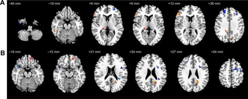
Table 5 Altered resting-state functional connectivity regions of the left cerebellum posterior lobe after sleep deprivation versus normal sleep

Table 6 Altered resting-state functional connectivity regions of the right cerebellum posterior lobe after sleep deprivation versus normal sleep