Figures & data
Figure 1 CT and CTA images before treatment.
Notes: (A) CT axial scanning reveals a hyperdensity mass on the left of sella turcica. (B) CT contrast scanning reveals enhancement of the parasella mass and dilatation and tortuosity of the left superior ophthalmic vein. (C) CTA image of anterior–posterior projection reveals an enhanced mass at the posterior wall of ICA and enlarged ophthalmic vein. (D) CTA reconstruction image reveals an enhanced mass at the lateral wall of left ICA-associated CCF and the drainage vein.
Abbreviations: CT, computed tomography; CTA, computed tomography angiography; ICA, internal carotid artery; CCF, carotid-cavernous fistula.
Abbreviations: CT, computed tomography; CTA, computed tomography angiography; ICA, internal carotid artery; CCF, carotid-cavernous fistula.
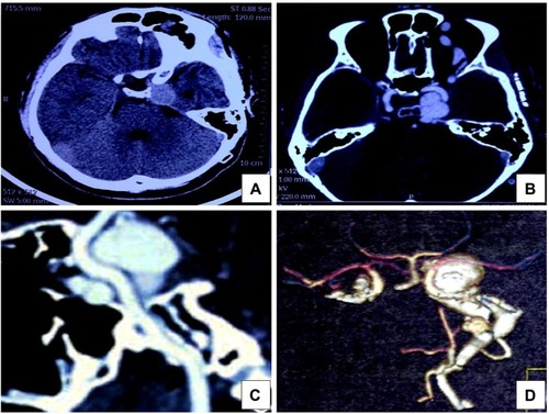
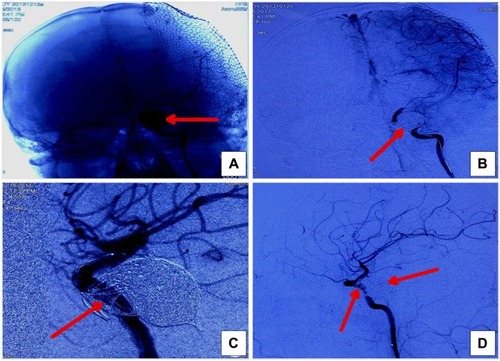
Figure 2 DSA images before treatment.
Notes: (A) Frontal and (B) lateral angiographic projection of left ICA demonstrates CCF. (C) Microcatheter enters the pouch of the cavernous sinus through the supraclinoid ICA and the PComA. (D) Three-dimensional angiography demonstrates the relationship between the PComA and CCF.
Abbreviations: DSA, digital subtraction angiography; ICA, internal carotid artery; CCF, carotid-cavernous fistula; PComA, posterior communicating artery.
Abbreviations: DSA, digital subtraction angiography; ICA, internal carotid artery; CCF, carotid-cavernous fistula; PComA, posterior communicating artery.

Figure 3 Post-embolization DSA images after endovascular embolization.
Notes: (A and B) Anterior–posterior projection. (C and D) Lateral projection. DSA of the left ICA reveals obliteration of the fistula and no residual filling of the CS in late venous phase. The red arrows indicate detachable platinum coils delivered into the pouch of the cavernous sinus.
Abbreviations: DSA, digital subtraction angiography; ICA, internal carotid artery; CS, cavernous sinus.
Abbreviations: DSA, digital subtraction angiography; ICA, internal carotid artery; CS, cavernous sinus.