Figures & data
Figure 1 Scheme of stabilization of GCC using protein G.
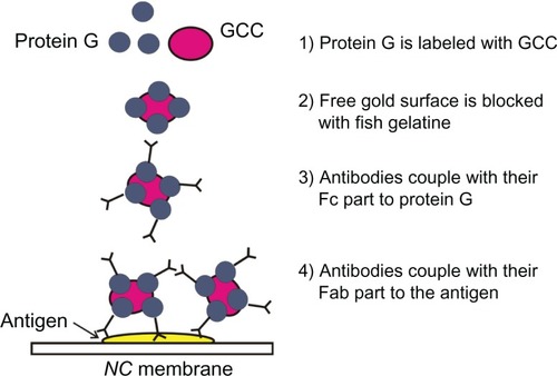
Figure 2 Color of GCC solution. A) after the addition of 1 μL. B) after the addition of 10 μL. C) pc-anti-BSA Ab to 10 mL of GCC solution, GCC flocculates after the addition of 1 μL of pc-antiperoxidase Ab to 10 mL of GCC solution, and its red color turns to violet and then blue. D) black color by total precipitation of GCC solution. E) GCC solution after the addition of fish gelatin. F) TEM image of GCC in A. G) TEM image of C. H) TEM image of D (I).
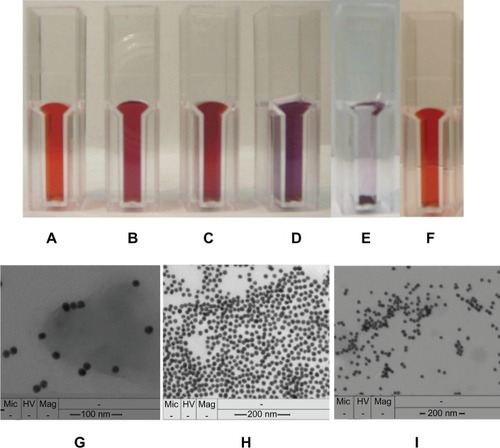
Table 1 The solutions used to produce labeled-GCC-Ab including protein G
Figure 3 Detection of 1 μL of HSA (200 mg/mL) dotted onto NC membrane after incubation in GCC (10 mL), including different amounts (B, 10 μL; D, 1 μL) of labeled mc-anti-HSA Ab. Images of these GCC-labeled Ab solutions (A, 10 μL; C, 1 μL) were checked by microscopy (Olympus).
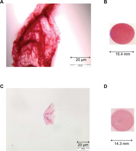
Figure 4 A) CLISA test: wells of microtiter plates were coated with HSA samples and incubated in the following:
A: GCC labeled with mc-anti-HSA Ab including GCC-blocking solution
B: GCC solution including GCC-blocking solution (negative control)
C: GCC labeled with mc-anti-β-galactosidase Ab including GCC-blocking solution (negative control)
D: GCC labeled with mc-anti-HSA Ab, including GCC-blocking solution, incubated in blocked, washed, and without coated HSA-sample wells (negative control)
E: GCC labeled with pc-anti-BSA Ab including GCC-blocking solution (for testing the specificity of Ab)
F: incubation of GCC solution (without GCC-blocking solution) in wells coated with samples (to show the importance of blocking GCC particles using GCC-blocking solution)
Figure 4 B) ELISA test, incubated in:
A: wells of microtiter plates were coated with tris or HCl buffer and incubated in mc-anti-HSA Ab solution (negative control)
B: wells of microtiter plates were coated with HSA samples and incubated in mc-anti-HSA Ab solution.
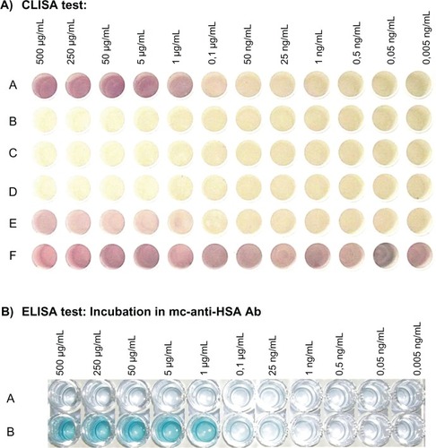
Figure 5 GCC flocculation can be avoided by increasing the amount of GCC solution, which was incubated with pc-antiperoxidase Ab to produce GCC-labeled Ab. A) GCC flocculation can be avoided by addition of protein G to the GCC solution before incubation with the Ab. B) No color reaction was observed by dotted β-LG protein (negative control).
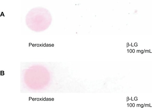
Figure 6 Positive signal of detected β-LG using the GCC labeling method on NC membranes.
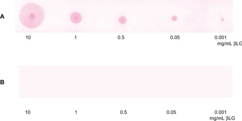
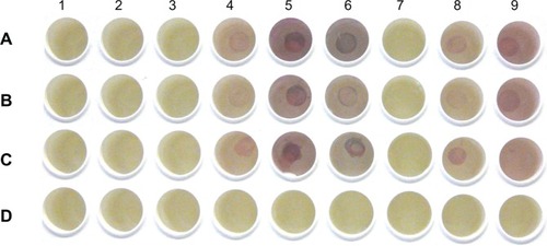
Figure 7 Detection of IgE in patients’ sera shows cherry red color signals. Columns: allergens: 1, 044 fungi I (380 μg protein/mL); 2, 106 mugwort (standardized concerning biological activity); 3, 154 short ragweed (320 μg/mL); 4, 015 grasses/cereals (630 μg/mL); 5, 708 Dermatophagoides farinae (130 μg/mL; dust mite, Ed.); 6, 725 Dermatophagoides pteronyssinus (standardized concerning biological activity; house dust mite, Ed.); 7, 108 birch tree (standardized for biological activity); 8, 116 ash tree (320 μg/mL); 9, GCC solution including GCC-blocking solution as a negative control – see section Results and Discussion. Wells of rows A, B, and C were incubated with patients’serum. Wells of row D were blocked, washed, and used as negative control.