Figures & data
Figure 1 Many nanoparticles have been investigated for biomedical applications targeting cancer.
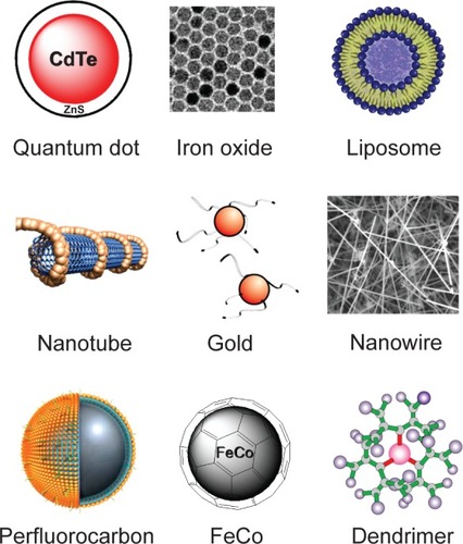
Figure 2 Different types of gold nanoparticles.
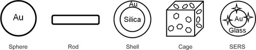
Figure 3 Gold nanoparticles have been modified with various molecules for in vitro assays.

Figure 4 Gold nanoparticles have been investigated for cell and phantom imaging using various techniques. Adapted from Chen et al 2005; Yang et al 2005; de la Fuente et al 2006; Durr et al 2007; Li et al 2007a; Oyelere et al 2007. Scale bar: 1 mm.
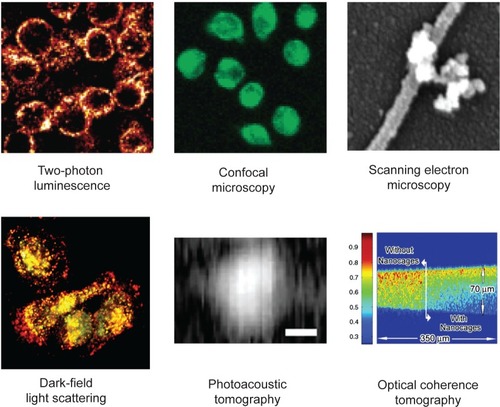
Figure 5 Multiplexed in vivo Raman imaging using SERS nanoparticles. Copyright © 2008, PNAS. Adapted with permission from Keren S, Zavaleta C, Cheng Z, et al. 2008. Noninvasive molecular imaging of small living subjects using Raman spectroscopy. Proc Natl Acad Sci U S A, 105:5844–9.
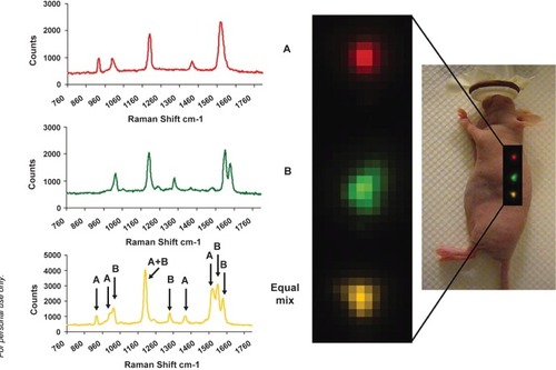
Figure 6 Gold nanoshells can destroy cancer cells both in vitro and in vivo. a. Cells incubated with gold nanoshells can be killed by NIR light (dark area). b. Temporal plots of maximum temperature change of NIR-irradiated tumors with and without nanoshells at depths of 2.5 mm and 7.3 mm beneath the tissue surface. c. Gross pathology after in vivo treatment with nanoshells and NIR laser revealed hemorrhaging and loss of tissue birefringence beneath the apical tissue surface. Hematoxylin/eosin (H&E) staining within the same plane confirms tissue damage within the area that contains nanoshells. Copyright © 2008, PNAS. Adapted with permission from Hirsch LR, Stafford RJ, Bankson JA, et al. 2003b. Nanoshell-mediated near-infrared thermal therapy of tumors under magnetic resonance guidance. Proc Natl Acad Sci U S A, 100:13549–54.
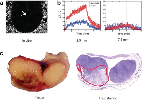
Figure 7 The versatile properties of gold nanoparticles have been employed for biomedical applications in many areas.
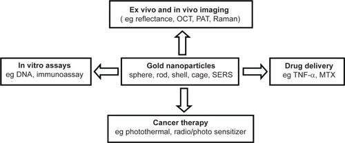
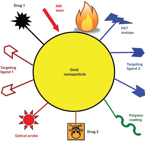
Figure 8 A multifunctional gold nanoparticle-based platform incorporating multiple receptor targeting, multimodality imaging, and multiple therapeutic entities. Not all functional moieties will be necessary and only suitably selected components are needed for each individual application.