Figures & data
Table 1 Demographic, biophysical, and sleep data
Table 2 Effects of race on airway dimensions
Table 3 Area and length effects of OSA with increasing severity vs control subjects
Figure 1 Reduced epiglottis cross-sectional area in an OSA subject over a control subject.
Notes: Each pair of MRI scans shows images obtained from the level of the dorsal tip of the epiglottis during wakefulness. Control subject (A) midsagittal MRI image of airway, and (B) axial image at the epiglottis level. Measures of cross-sectional area were taken at the horizontal white lines in images (A, C). OSA subject (C) midsagittal MRI image shows narrowing (arrow) of pharyngeal upper airway at an elongated soft palate, and (D) axial image shows representative reduced cross-sectional area at the epiglottis level. The cross-sectional areas corresponding to the horizontal lines are outlined in tracings (B, D).
Abbreviations: OSA, obstructive sleep apnea; MRI, magnetic resonance imaging.
Abbreviations: OSA, obstructive sleep apnea; MRI, magnetic resonance imaging.
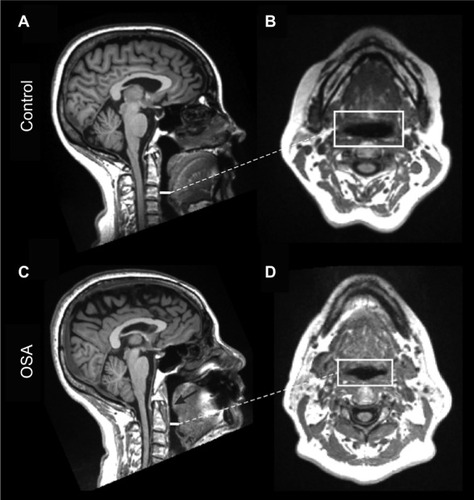
Figure 2 Increased oropharyngeal length in OSA subjects with differing severity vs a control subject.
Notes: Midsagittal MRI of oropharyngeal airway length during wakefulness, measured as the distance from the hard palate to the dorsal tip of the epiglottis, of a female control subject (A), female OSA subject with moderate AHI (B), and female OSA subject with severe AHI (C).
Abbreviations: OSA, obstructive sleep apnea; MRI, magnetic resonance imaging; AHI, apnea–hypopnea index.
Abbreviations: OSA, obstructive sleep apnea; MRI, magnetic resonance imaging; AHI, apnea–hypopnea index.
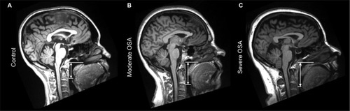
Table 4 Area and length effects of sex and OSA
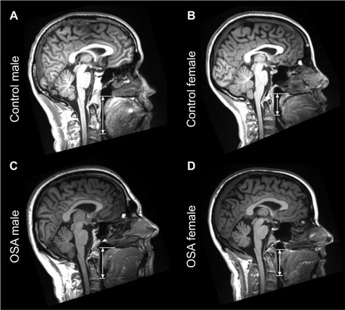
Figure 3 Increased oropharyngeal length in a male control and OSA subject vs a female control and OSA subject.
Notes: Midsagittal MRI of the OPAL of a male control subject (A) compared to a female control subject (B), and a male OSA subject (C) compared to a female OSA subject (D). Males displayed elongated OPAL over females.
Abbreviations: OSA, obstructive sleep apnea; MRI, magnetic resonance imaging; OPAL, oropharyngeal airway length.
Abbreviations: OSA, obstructive sleep apnea; MRI, magnetic resonance imaging; OPAL, oropharyngeal airway length.